Page 1 of 9
AN10.1-13 | Axilla, Shoulder and Scapular region — SDL Guide
Learning Objectives
- Describe the boundaries and contents of the axilla, including axillary artery, vein, and lymph nodes (AN10.1, AN10.2, AN10.4, AN10.7)
- Trace the formation of the brachial plexus and explain the clinical significance of Erb's palsy and Klumpke's paralysis (AN10.3, AN10.5, AN10.6)
- Describe the muscles acting on the shoulder joint — trapezius, latissimus dorsi, rotator cuff (SITS), and serratus anterior (AN10.8, AN10.9, AN10.10, AN10.12)
- Explain the anatomy of the shoulder joint, its movements, and the scapular anastomosis (AN10.11, AN10.13)
- Identify the clinical consequences of axillary nerve injury and understand safe intramuscular injection technique (AN10.12)
INSTRUCTIONS
This module covers the axilla (armpit), brachial plexus (the nerve highway to your arm), and shoulder region. We'll start from what you can feel on your own body and build from there.
Parallel connections: In Physiology, you're learning about nerve conduction and muscle contraction — the same mechanisms that power shoulder movements. In Biochemistry, you're studying extracellular matrix proteins (collagen, elastin) — these form the tendons and ligaments that hold the shoulder joint together.
References
- OpenStax Anatomy and Physiology 2e, Chapter 11: The Muscular System (textbook (CC BY 4.0))
- B.D. Chaurasia's Human Anatomy, Vol. 1, Ch 5-7: Pectoral Region, Axilla, Shoulder (textbook)
- Netter's Atlas of Human Anatomy, 8th ed., Plates 408–423 (Shoulder and Axilla) (atlas)
- Gray's Anatomy for Students, 4th ed., Chapter 7: Upper Limb (textbook)
Version 2.0 | NMC CBUC 2024, Adapted from OpenStax A&P 2e (CC BY 4.0)
CLINICAL SCENARIO
Raise your arm and look at your armpit. It seems like a simple fold of skin — but hiding beneath that skin is one of the most important crossroads in your body. Every major blood vessel, nerve, and lymphatic channel travelling to your arm passes through this narrow space. Surgeons approach it with great respect during breast cancer surgery, because a single slip can paralyse the arm. This crowded passageway has a name: the axilla — and understanding it is your gateway to the entire upper limb.
WHY THIS MATTERS
As a doctor, the axilla matters to you in three ways. First, during a breast examination, you'll palpate axillary lymph nodes — the most common site where breast cancer spreads. Second, in the emergency department, axillary artery injuries from shoulder dislocations need urgent recognition. Third, every time you trace a nerve to the arm (brachial plexus), you'll pass through the axilla. Think of it as the Grand Central Station of the upper limb — everything passes through here.
RECALL
From your earlier study of the body wall, you know that the pectoralis major is the large chest muscle (the one you feel when you push against something). You also know that muscles have origins (fixed attachment) and insertions (moving attachment). We'll build on these concepts as we explore the axilla's walls.
The Axilla — A Pyramid-Shaped Space (AN10.1)
The axilla is not just the surface 'armpit' — it's a pyramid-shaped space between the chest wall and the upper arm. Like any pyramid, it has an apex (top), a base (bottom), and four walls.

Figure: The Axilla — A Pyramid-Shaped Space (AN10.1)
Imagine you're looking at a cross-section through someone's armpit:
- Apex — the narrow top of the pyramid, formed by the gap between the clavicle (front), scapula (back), and first rib (medially). This is the gateway through which vessels and nerves enter the axilla from the neck. It's called the cervicoaxillary canal.
- Base — the floor, formed by the skin and axillary fascia (the concave skin of the armpit you can see).
- Anterior wall — the front, formed by the pectoralis major and pectoralis minor muscles. These are the muscles you feel on your chest.
- Posterior wall — the back, formed by the subscapularis, latissimus dorsi, and teres major muscles.
- Medial wall — the inner side, formed by the upper ribs and serratus anterior muscle (the finger-like muscle on the side of the chest).
- Lateral wall — the narrow outer side, formed by the intertubercular sulcus (bicipital groove) of the humerus.
Think of it this way: the axilla is like a pyramid tent. The chest muscles form the front flap, the back muscles form the rear, the ribs are one side, and the arm bone is the other. Everything going to the arm must squeeze through this tent.
The Axillary Artery — Three Parts and Their Branches (AN10.4)
The main blood supply to the upper limb enters through the apex of the axilla as the axillary artery — a continuation of the subclavian artery that changes its name as it crosses the outer border of the first rib.

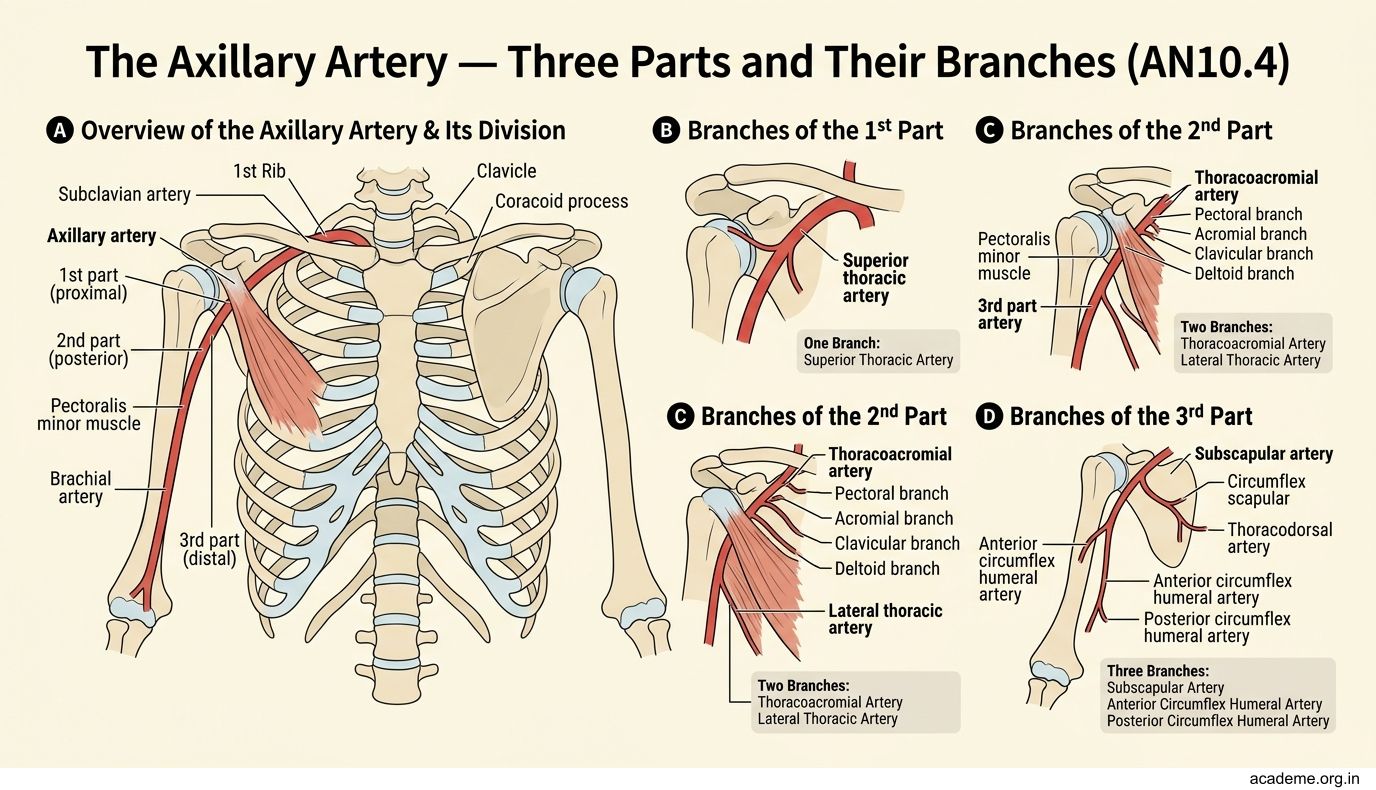
Figure: The Axillary Artery — Three Parts and Their Branches (AN10.4)
The pectoralis minor muscle crosses the axillary artery and divides it into three parts (this is a favourite exam question!):
- 1st part — above (proximal to) pectoralis minor → gives off 1 branch: the superior thoracic artery
- 2nd part — behind pectoralis minor → gives off 2 branches: the thoracoacromial artery and the lateral thoracic artery
- 3rd part — below (distal to) pectoralis minor → gives off 3 branches: the subscapular artery, the anterior circumflex humeral artery, and the posterior circumflex humeral artery
Memory trick: The number of branches matches the part number — 1, 2, 3 branches for the 1st, 2nd, and 3rd parts respectively. Simple!
The axillary artery becomes the brachial artery at the lower border of teres major — which is the artery you feel when taking blood pressure.
Axillary Lymph Nodes — The Sentinel Guards (AN10.7)
The axilla contains about 20–30 lymph nodes arranged in groups. These nodes filter lymph from the upper limb, breast, and chest wall — making them critically important in breast cancer staging.

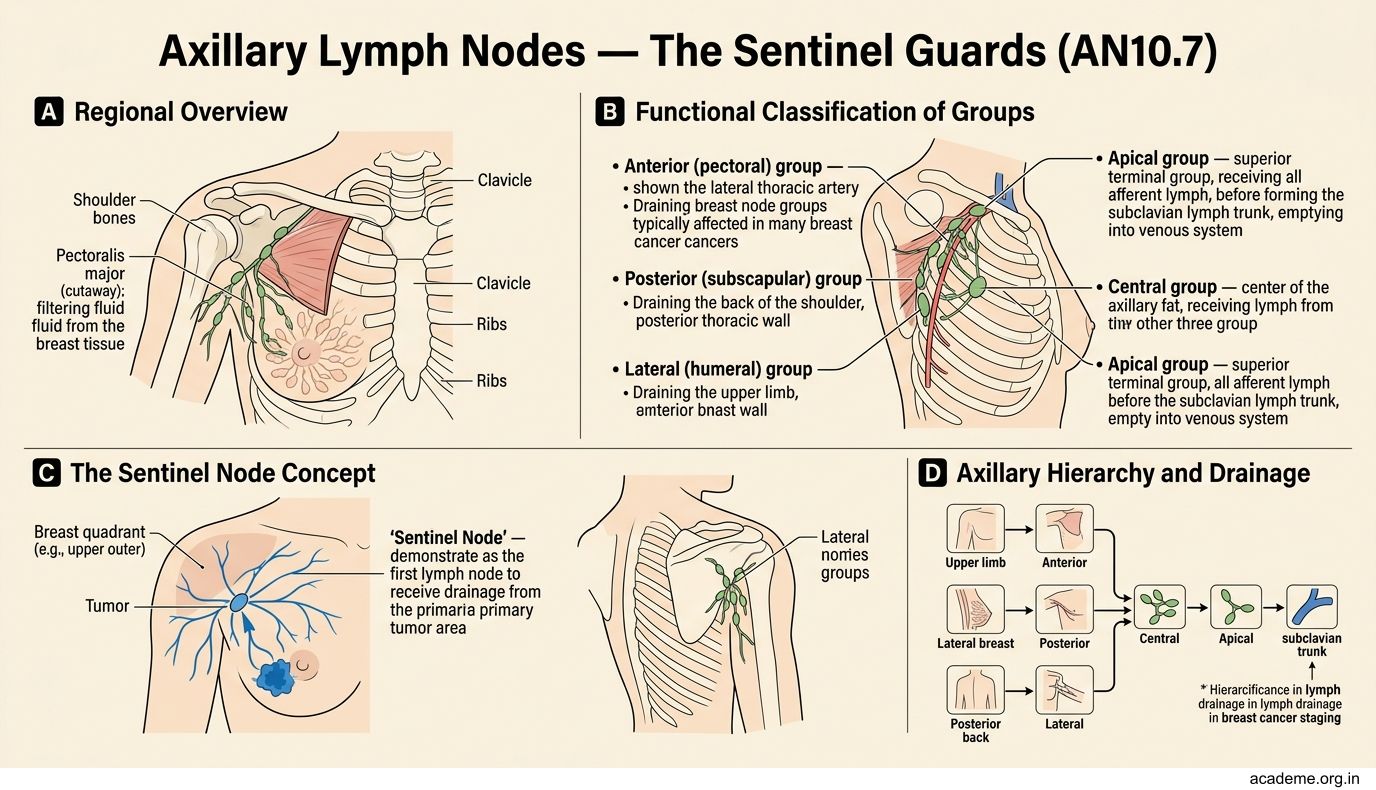
Figure: Axillary Lymph Nodes — The Sentinel Guards (AN10.7)
The groups are named by their location within the axilla:
- Anterior (pectoral) group — along the lateral thoracic artery, drains the breast and anterior chest wall. These are the first nodes to receive breast cancer metastasis.
- Posterior (subscapular) group — along the subscapular artery, drains the back and posterior shoulder.
- Lateral group — along the axillary vein, drains the upper limb.
- Central group — in the fat of the axilla, receives lymph from all three groups above.
- Apical group — at the apex of the axilla, receives lymph from the central group and drains into the subclavian lymph trunk.
Clinical connection: During breast cancer surgery, surgeons perform a sentinel lymph node biopsy — they inject a dye near the tumour and trace which lymph node it reaches first (the 'sentinel' node). If that node is cancer-free, the remaining nodes are likely clear too, avoiding unnecessary removal of all axillary nodes (which can cause arm swelling called lymphoedema).
Contents of the Axilla — What's Inside the Pyramid (AN10.2)
Now that you know the walls and lymph nodes, here's the complete inventory of what's packed inside the axilla:
- Axillary artery and its branches (the blood supply highway going to the arm)
- Axillary vein and its tributaries (the return highway, lying medial to the artery)
- Brachial plexus — the network of nerves supplying the entire upper limb (we'll cover this in Part 2)
- Axillary lymph nodes — 5 groups as described above
- Axillary fat — filling the spaces between these structures
- Axillary tail of the breast — a projection of breast tissue that extends into the axilla (important: lumps here can be breast tissue, not just lymph nodes)
All these structures are wrapped in the axillary sheath, an extension of the prevertebral fascia from the neck that accompanies the vessels and nerves through the cervicoaxillary canal.
SELF-CHECK
The axillary artery is divided into three parts by a muscle crossing it. Which muscle is this, and how many branches does the 2nd part give off?
A. Pectoralis major; 2 branches
B. Pectoralis minor; 2 branches
C. Pectoralis minor; 3 branches
D. Subscapularis; 1 branch
Reveal Answer
Answer: B. Pectoralis minor; 2 branches
The pectoralis minor crosses the axillary artery and divides it into three parts. The 2nd part (behind pectoralis minor) gives off 2 branches: the thoracoacromial artery and the lateral thoracic artery. Remember: the number of branches matches the part number — 1, 2, 3.