Page 2 of 6
AN12.1-15 | Forearm & hand — SDL Guide (Part 2)
Part 4: Dorsal Forearm — Muscles, Nerves, Vessels, and Wrist Drop
The Posterior (Extensor) Compartment (AN12.11)

Figure: Wrist Drop (AN12.13)

Figure: Nerves and Vessels of the Back of the Forearm (AN12.12)

Figure: Part 4: Dorsal Forearm — Muscles, Nerves, Vessels, and Wrist Drop
The posterior forearm contains 12 muscles in two layers, all supplied by the posterior interosseous nerve (PIN = deep branch of radial nerve), EXCEPT brachioradialis and extensor carpi radialis longus (ECRL) — which are supplied by the radial nerve proper.
Superficial Layer (7 muscles, from lateral epicondyle):
Brachioradialis, ECRL, ECRB (extensor carpi radialis brevis), extensor digitorum (ED), extensor digiti minimi (EDM), extensor carpi ulnaris (ECU), anconeus.
Deep Layer (5 muscles, from posterior radius/ulna/interosseous membrane):
Supinator, abductor pollicis longus (APL), extensor pollicis brevis (EPB), extensor pollicis longus (EPL), extensor indicis (EI).
Nerves and Vessels of the Back of the Forearm (AN12.12)
Posterior interosseous nerve (PIN):
• Deep branch of the radial nerve
• Enters the posterior compartment by passing through the supinator muscle (winding around the lateral neck of radius)
• Supplies ALL posterior compartment muscles except brachioradialis + ECRL
• Terminates as the posterior interosseous nerve of the wrist (sensory to dorsal wrist joint)
Compression at the supinator → radial tunnel syndrome (pain without sensory loss) or PIN palsy (finger drop without wrist drop, since ECRL intact).
Superficial radial nerve: Purely sensory; runs under brachioradialis; supplies dorsal lateral hand and proximal 3½ fingers (dorsal).
Wrist Drop (AN12.13)
Wrist drop = radial nerve injury in the spiral groove of the humerus (mid-shaft fracture).
Muscles paralysed: All wrist extensors (ECRL, ECRB, ECU), extensor digitorum, EDM, EI, APL, EPL, EPB.
Triceps often spared (its nerve branches arise proximal to the spiral groove).
| Level of injury | Wrist drop | Triceps | Finger drop |
|---|---|---|---|
| Axilla | Yes | Yes (paralysed) | Yes |
| Spiral groove | Yes | Usually spared | Yes |
| Lateral epicondyle (PIN) | No | Spared | Yes |
Mnemonic: BEST muscles lost in wrist drop = Brachioradialis, Extensors, Supinator, Triceps (sometimes).
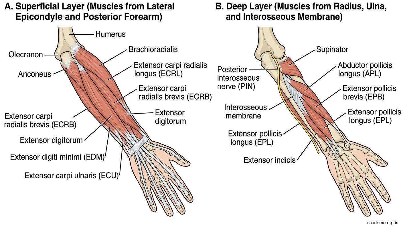
Figure: Part 4: Dorsal Forearm — Muscles, Nerves, Vessels, and Wrist Drop
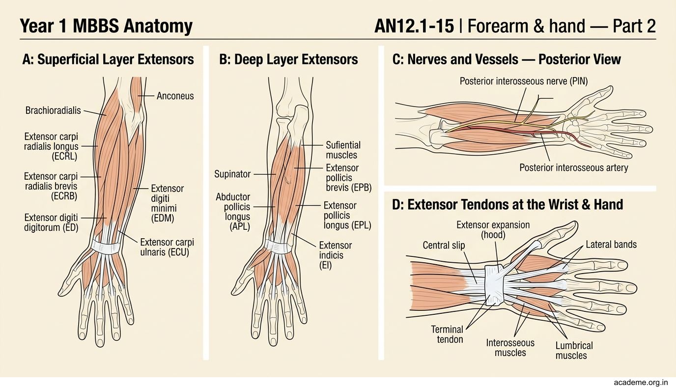
Figure: Nerves and Vessels of the Back of the Forearm (AN12.12)
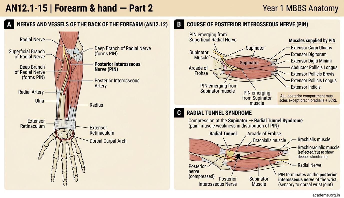
Figure: Wrist Drop (AN12.13)
CLINICAL PEARL
Saturday night palsy is a classic cause of radial nerve injury at the spiral groove. It occurs when a person falls asleep with their arm draped over the back of a chair (also called "crutch palsy" when axillary crutches compress the nerve). The patient wakes with wrist drop and sensory loss on the dorsum of the hand.
In India, this presentation is seen after heavy alcohol intoxication or post-operative prolonged positioning. Always examine the triceps reflex — if it is preserved, the injury is at or below the spiral groove (not in the axilla).
SELF-CHECK — : Dorsal Forearm & Wrist Drop
A patient has wrist drop with preserved triceps function and normal sensation over the arm. Where is the radial nerve most likely injured?
A. In the axilla
B. In the spiral groove of the humerus
C. At the lateral epicondyle
D. As the PIN in the supinator
Reveal Answer
Answer: B. In the spiral groove of the humerus
Which posterior forearm muscle is NOT supplied by the posterior interosseous nerve?
A. Extensor carpi ulnaris
B. Extensor carpi radialis longus
C. Extensor digitorum
D. Supinator
Reveal Answer
Answer: B. Extensor carpi radialis longus
Part 5: Extensor Retinaculum, Anatomical Snuffbox, and Extensor Expansion
The Extensor Retinaculum (AN12.14)

Figure: Extensor Expansion (Dorsal Digital Expansion) (AN12.15)

Figure: The Anatomical Snuffbox

Figure: Part 5: Extensor Retinaculum, Anatomical Snuffbox, and Extensor Expansion
The extensor retinaculum is a thickened band of deep fascia on the dorsum of the wrist, running from the lateral radius to the triquetrum and pisiform medially. It sends vertical septa creating 6 osseofibrous compartments:
| Compartment | Tendons | Clinical note |
|---|---|---|
| 1 (lateral) | APL + EPB | De Quervain's tenosynovitis here |
| 2 | ECRL + ECRB | — |
| 3 | EPL | Winds around Lister's tubercle |
| 4 | ED + EI | — |
| 5 | EDM | — |
| 6 (medial) | ECU | — |
De Quervain's tenosynovitis: Inflammation of the 1st compartment (APL + EPB). Pain at the radial wrist aggravated by thumb movement. Positive Finkelstein's test (ulnar deviation of wrist while thumb is flexed across palm).
The Anatomical Snuffbox
The anatomical snuffbox is a triangular depression visible on the dorsolateral wrist when the thumb is extended and abducted.
- Medial (posterior) boundary: Tendon of EPL
- Lateral (anterior) boundary: Tendons of APL and EPB
- Floor: Scaphoid and trapezium (also the radial artery and branches of the superficial radial nerve)
- Roof: Superficial radial nerve, cephalic vein
Clinical importance: Tenderness in the anatomical snuffbox after a FOOSH injury = suspect scaphoid fracture even with a normal X-ray. The radial artery passes through the floor to reach the palm and form the deep palmar arch.
Extensor Expansion (Dorsal Digital Expansion) (AN12.15)
The extensor expansion (extensor hood) is a triangular fibrous aponeurosis on the dorsum of each finger.
Formation (mnemonic: ELI):
• E = Extensor digitorum tendon (fans out over dorsum of proximal phalanx)
• L = Lumbricals (contribute via lateral bands on each side)
• I = Interossei (contribute via lateral bands on each side)
Insertions:
• Central slip → base of middle phalanx
• Lateral bands (convergence of lumbrical + interosseous contributions + ED) → base of distal phalanx
Pathology:
• Boutonnière deformity: Rupture of the central slip → middle phalanx herniates through lateral bands → PIPJ flexed + DIPJ extended
• Swan-neck deformity: Volar plate laxity at PIPJ → lateral bands shift dorsally → PIPJ hyperextended + DIPJ flexed
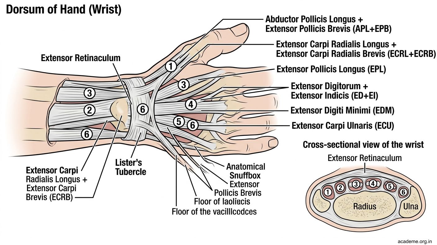
Figure: Part 5: Extensor Retinaculum, Anatomical Snuffbox, and Extensor Expansion
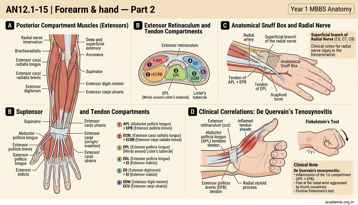
Figure: The Anatomical Snuffbox
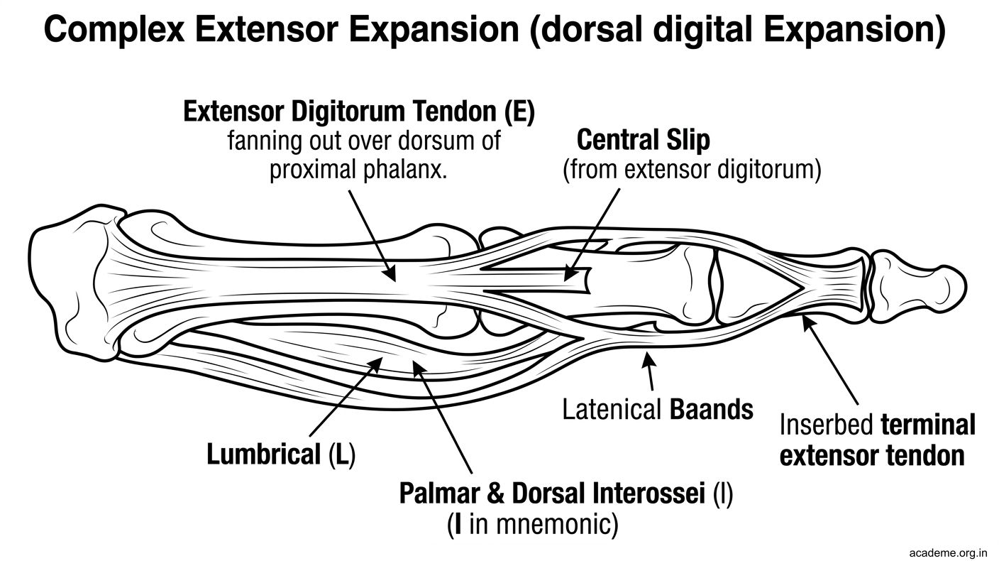
Figure: Extensor Expansion (Dorsal Digital Expansion) (AN12.15)
SELF-CHECK — : Extensor Retinaculum & Expansion
Which tendon forms the MEDIAL boundary of the anatomical snuffbox?
A. Extensor pollicis brevis
B. Abductor pollicis longus
C. Extensor pollicis longus
D. Extensor carpi radialis longus
Reveal Answer
Answer: C. Extensor pollicis longus
The extensor expansion receives contributions from which muscles on the lateral sides (the lateral bands)?
A. FDS and FDP
B. Lumbricals and interossei
C. Extensor digitorum and brachioradialis
D. Dorsal interossei only
Reveal Answer
Answer: B. Lumbricals and interossei