Page 1 of 7
AN4.1-5 | General features of skin and fascia — SDL Guide
Learning Objectives
- Describe the two main types of skin (thick and thin) and explain how dermatomes map sensation
- Identify the layers and appendages of skin using a cross-section diagram
- Describe superficial fascia, its contents, and how fat distribution varies across the body
- Describe deep fascia, its modifications, and their clinical functions
- Explain why the direction of a surgical incision matters (Langer's lines)
INSTRUCTIONS
This is one of the earliest topics in your anatomy journey — we'll start from what you can see and touch on your own body. No dissection room knowledge needed yet.
Parallel connections: In Biochemistry, you're learning about proteins — the same collagen and keratin proteins that give skin its strength. In Physiology, you're studying general physiology — how cells communicate and maintain boundaries, which is exactly what skin does for the whole body.
References
- OpenStax Anatomy and Physiology 2e, Chapter 5: The Integumentary System (textbook (CC BY 4.0))
- B.D. Chaurasia's Human Anatomy, Vol. 1, Chapter 1: Introduction (textbook)
- Netter's Atlas of Human Anatomy, 8th ed., Plates 1–4 (Skin and Fascia) (atlas)
Version 2.0 | NMC CBUC 2024, Adapted from OpenStax A&P 2e (CC BY 4.0)
CLINICAL SCENARIO
Your skin is the largest organ in your body — it weighs about 3.5 kg and covers approximately 2 square metres. That's bigger than a large bath towel. Right now, as you read this, your skin is doing at least five jobs simultaneously: keeping water in, keeping bacteria out, sensing temperature, making vitamin D from sunlight, and regulating your body temperature. And here's something most people don't realise — if you pinch the skin on the back of your hand, it springs back. Pinch the skin on your elbow, and it stays pinched for a moment. Why? The answer lies in how skin is built — and that's what we'll learn today.
WHY THIS MATTERS
As a doctor, you will examine skin every single day — it's the first thing you see on a patient. You'll need to know why a rash spreads in a particular pattern (dermatomes), why a surgeon cuts in one direction and not another (Langer's lines), and why injections are given in certain spots (fat distribution in superficial fascia). Skin is also the organ where you'll first learn to read disease — from the yellow skin of jaundice to the bluish tinge of cyanosis.
RECALL
From school biology, you know that skin is made of layers — you've probably heard the terms epidermis (outer layer) and dermis (inner layer). You know skin has hair, sweat glands, and nails. You also know that skin acts as a barrier against germs. Let's build on this: we'll go from 'skin has layers' to understanding exactly what each layer does and why it matters clinically.
Two Types of Skin (AN4.1)
Look at your palm. Now look at the back of your hand. They feel different, don't they? That's because your body has two types of skin:
- Thick skin — found on the palms of your hands and soles of your feet. It has a very thick epidermis (up to 1.5 mm) with five distinct layers. It has no hair follicles but has many sweat glands. The thick epidermis protects against friction and wear — think of how much your palms and soles are used.
- Thin skin — covers the rest of your body. It has a thinner epidermis (about 0.1 mm) with fewer layers. It has hair follicles, sebaceous (oil) glands, and sweat glands.
Despite the names, thin skin is NOT fragile — it's simply more flexible. The skin on your eyelids (the thinnest skin in the body) is delicate, but the skin on your back (also thin skin) is quite tough.
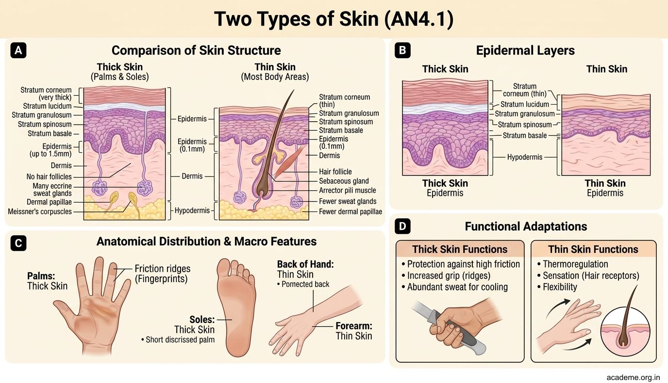

Figure: Two Types of Skin (AN4.1)
Dermatomes — A Map of Skin Sensation
Here's a fascinating fact: your skin is divided into zones, each supplied by a single spinal nerve. These zones are called dermatomes (from Greek: derma = skin, tome = cutting).
Imagine your body painted in coloured stripes, each stripe representing the area of skin supplied by one nerve. On the trunk, these stripes wrap horizontally around your body (like a stack of rings). On the limbs, they run lengthwise.
Why do dermatomes matter? Because they create a map that doctors use every day:
- If a patient has numbness in a specific strip of skin, you can identify which nerve is damaged
- If a patient has shingles (a reactivation of the chickenpox virus), the rash follows one dermatome exactly — because the virus lives in one nerve root
- Anaesthetists use dermatome maps to check that a spinal block is working at the right level
You don't need to memorise the full map right now — we'll revisit dermatomes in detail during the nervous system (Week 32–37). For now, just understand the concept: one spinal nerve → one strip of skin.
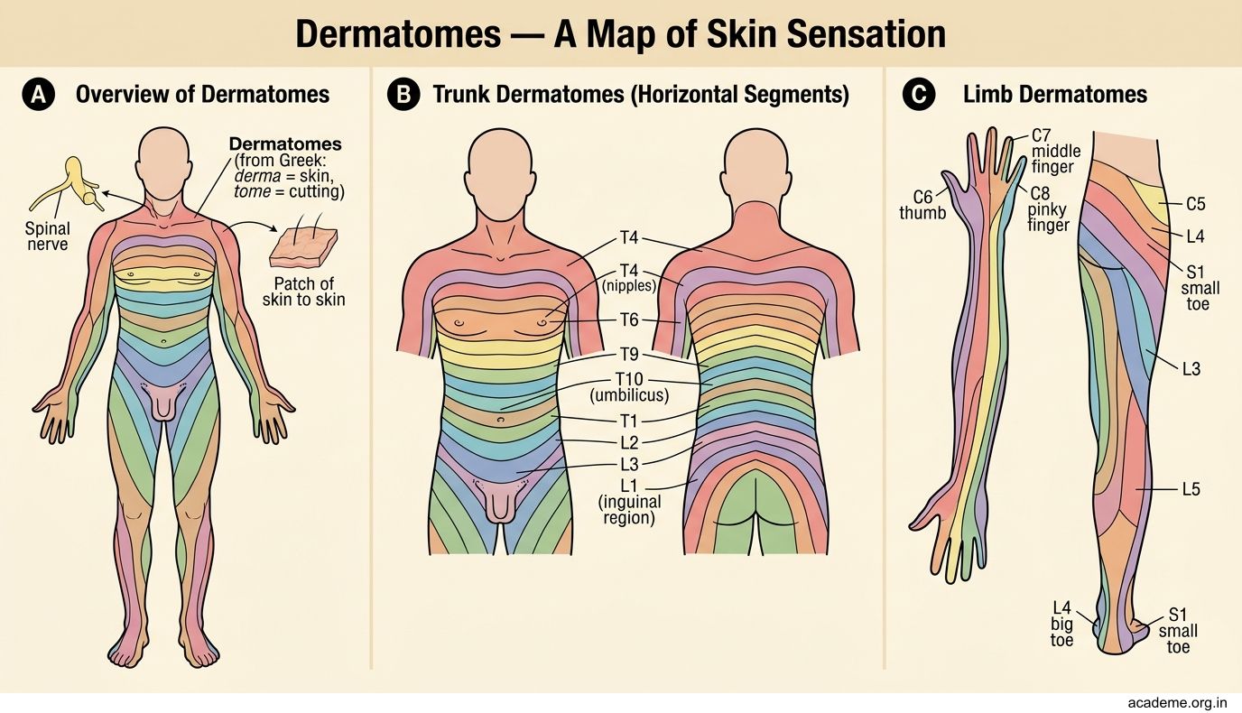

Figure: Dermatomes — A Map of Skin Sensation
SELF-CHECK
A patient presents with a painful, blistering rash that wraps around the left side of their chest in a band-like pattern, following one strip of skin precisely. This pattern corresponds to:
A. A blood vessel territory
B. A dermatome — the area supplied by one spinal nerve
C. A lymphatic drainage zone
D. A random distribution typical of allergic reactions
Reveal Answer
Answer: B. A dermatome — the area supplied by one spinal nerve
This is classic shingles (herpes zoster) — the virus reactivates in one nerve root and causes a rash limited to that nerve's dermatome. The band-like pattern following a single strip of skin is the clinical giveaway. This is why understanding dermatomes is essential for diagnosis.
Layers of the Skin — From Surface to Depth (AN4.2)
Skin has two main layers (you already know their names from school):
1. Epidermis — the outermost layer. It's made entirely of epithelial cells and contains no blood vessels (it's avascular). Nutrients reach it by diffusion from the layer below. The epidermis has several key features:
- Keratinocytes — the main cell type. They produce a tough protein called keratin that makes skin waterproof and resistant to wear. The same protein makes up your hair and nails.
- Melanocytes — cells that produce melanin, the pigment that gives skin its colour and protects against UV radiation. Everyone has roughly the same number of melanocytes — skin colour differences come from the amount and type of melanin produced.
- The outermost surface is made of dead, flattened keratinocytes that are constantly shed and replaced. Your entire epidermis replaces itself every 4–6 weeks.
2. Dermis — the thicker layer beneath the epidermis. Unlike the epidermis, the dermis is made of connective tissue and is rich in blood vessels, nerves, and glands. It contains:
- Collagen fibres — provide strength (this is the same collagen protein you're studying in Biochemistry right now)
- Elastic fibres — provide stretch and recoil (this is why young skin springs back when pinched)
- Hair follicles, sweat glands, sebaceous glands — the skin's appendages
- Sensory nerve endings — for touch, pressure, pain, and temperature
Below the dermis is the hypodermis (also called subcutaneous tissue or superficial fascia) — but this isn't technically part of the skin. We'll cover it next.
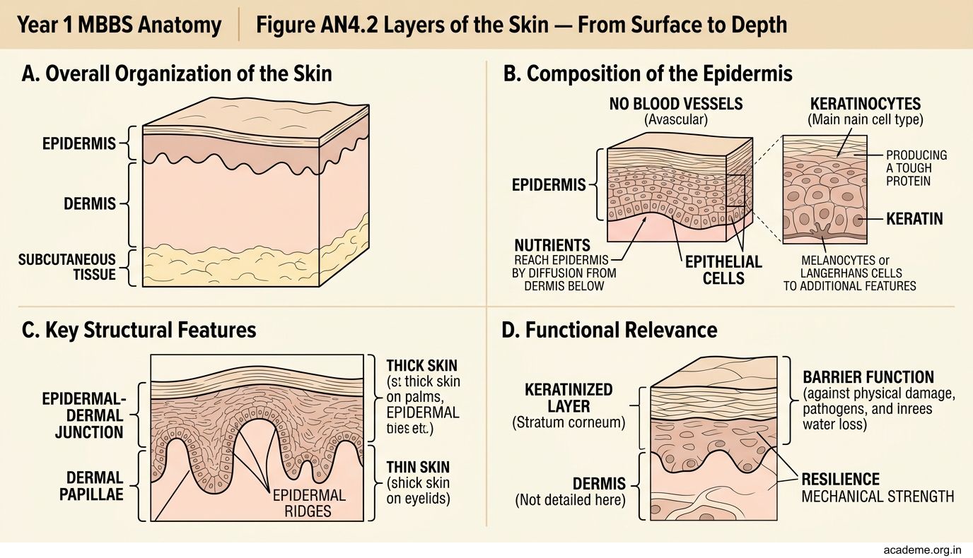

Figure: Layers of the Skin — From Surface to Depth (AN4.2)