Page 5 of 15
FM4.2-3,FM14.4 | Age & Dental Identification & Age Estimation — SDL Guide (Part 2)
Gustafson's Method for Adult Age Estimation from Teeth
Once all permanent teeth are fully erupted (after age 21), dental eruption patterns are no longer useful for age estimation. Gustafson's method (1950, modified for Indian populations) remains the most widely used and validated technique for estimating age from teeth in adults. It is based on the progressive, age-dependent changes that all human teeth undergo throughout adult life, which can be assessed histologically or macroscopically in extracted or sectioned teeth.
Gustafson identified six criteria that change progressively with age, each scored on a 4-point scale: 0 (absent/none), 1 (slight), 2 (moderate), 3 (marked/advanced):
- Attrition (A) — wearing away of the crown surface due to mastication; begins at occlusal surfaces; moderate by 40s, marked by 60s
- Secondary dentine deposition (S) — progressive narrowing of the pulp cavity as secondary dentine is deposited from the pulp wall inward; assessed on longitudinal section
- Cementum apposition (C) — incremental deposition of cementum on the root surface throughout life; assessed on cross-section at root apex
- Root transparency (R) — translucency of the root apex due to infilling of dentinal tubules with calcium salts; not visible macroscopically without transillumination; one of the most reliable criteria
- Root resorption (Re) — irregular resorption lacunae at the root apex; generally increases with age but highly variable
- Periodontal recession (P) — recession of the alveolar bone and supporting periodontal tissue, exposing the root; measured as the length of exposed root
The total score (sum of all 6 criteria, maximum 18) is entered into a regression formula. The regression formula derived from Gustafson's original work and validated with Indian population modifications (Reddy's) is: Estimated age = 11.43 + 4.56 × (total point score). The standard error of the estimate is ±3.63 years.
Practical example: If a tooth shows attrition score 2, secondary dentine 2, cementum 1, root transparency 2, root resorption 0, periodontal recession 1 → total = 8. Age estimate = 11.43 + (4.56 × 8) = 11.43 + 36.48 = 47.9 years → reported as '44–52 years (Gustafson's method, ±3.6 yr SE)'.
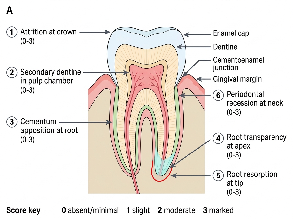
Gustafson Criteria for Dental Age Estimation
Limitations: Gustafson's method requires extracted or sectioned teeth, making it destructive and typically applicable to post-mortem cases. It cannot be applied to living persons without tooth extraction. For living persons, non-destructive modifications (radiographic root transparency, OPG-based pulp-to-tooth ratio assessment) are used. Additionally, pathological conditions (grinding disorder — bruxism causes accelerated attrition, making the person appear older than they are; periodontal disease accelerates recession) can lead to age overestimation.
Skeletal Age Estimation: Epiphyseal Fusion and Ossification
Skeletal age estimation uses the timing of epiphyseal fusion — the progressive ossification and closure of growth plates at the ends of long bones — as the primary biological clock for ages roughly 10–25 years. Radiological examination (X-ray of wrist, elbow, knee, pelvis, or clavicle depending on suspected age) is the standard investigative tool. The appearance of an epiphysis on X-ray as a separate radio-dense shadow, followed by progressive bridging and eventual fusion with the shaft, provides a sequence of datable events.
For medicolegal practice in India, the two most legally critical epiphyseal fusion sites are:
- Medial epiphysis of clavicle: the last epiphysis to fuse in the human body. It appears (~16–18 years) and fuses completely at approximately 25 years. Because its fusion period spans and extends beyond the critical 18-year threshold, the clavicle X-ray is the single most important radiological study in JJ Act age determination. A partially fused medial clavicular epiphysis places the subject between approximately 18–25 years.
- Iliac crest apophysis: appears approximately 14–15 years in females and 15–16 years in males; fuses completely at 20–21 years (Indian reference, Reddy's). A fused iliac crest indicates age >20–21 years.
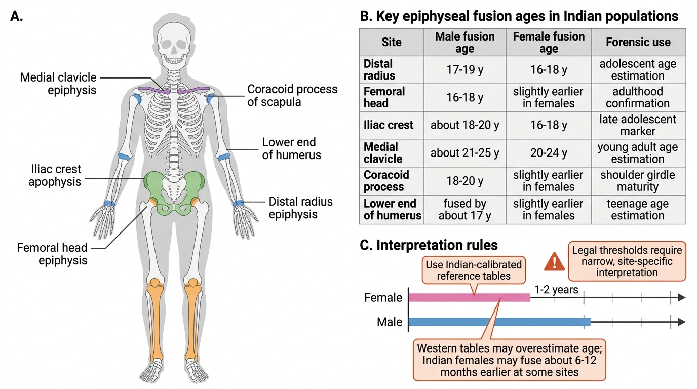
Epiphyseal Fusion Sites for Age Estimation in Indian Populations
Additional commonly assessed epiphyseal fusion sites and their approximate Indian fusion ages include:
- Distal radius: fuses ~17–19 years (male), 16–18 years (female) — used when adolescent age is in question
- Femoral head epiphysis: fuses ~16–18 years — helps confirm adulthood
- Coracoid process of scapula: fuses ~18–20 years
- Lower end of humerus: multiple separate centres, all fused by ~17 years
Important caveat: Indian population epiphyseal fusion data shows earlier fusion in some sites compared to Western data (by approximately 6–12 months for females), reinforcing the rule that Indian-calibrated tables must be used. Furthermore, sex-specific differences in fusion timing exist at most sites — females fuse earlier than males by 1–2 years on average — and the sex of the subject (from other parameters) should inform which reference table is used.
For degenerative skeletal markers used in older adults (above 30 years), pubic symphysis morphology (Todd's phases, Suchey-Brooks phases) and cranial suture closure provide approximate age ranges, but with wider error margins (±5–10 years) that are less useful in legal proceedings where narrow thresholds matter.
Bite Marks and Dental Record Matching
Beyond age estimation, forensic dentistry serves two additional identification functions: bite mark analysis and dental record matching. These are methods of individual (person-specific) identification rather than age estimation, completing the forensic dental toolkit.
Bite marks are patterned injuries inflicted by human or animal teeth, typically found on skin in cases of sexual assault, child abuse, homicide, or occasionally self-inflicted injury. The forensic value lies in the principle that each individual's dentition is unique — arch shape, spacing, individual tooth rotations, and wear patterns combine to create a dental signature. When a bite mark is found on a victim, its characteristics (arch shape, inter-canine distance, individual tooth imprints) can be compared with the dental impressions of a suspected perpetrator.
Documentation of bite marks follows a strict protocol: (1) photograph in situ with a scale ruler in two planes; (2) swab the bite area for salivary DNA before any other handling; (3) take impressions if the bite is on firm tissue (e.g., cheese, bread, bite on cadaver); (4) excise for histological examination if on a cadaver. The ABFO (American Board of Forensic Odontology) scale must be included in photographs to allow accurate dimensional comparison.
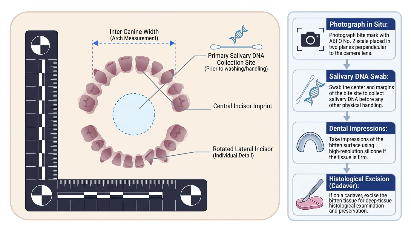
Provided image
Dental record matching is used to identify an unknown deceased person by comparing the teeth of the unidentified body with ante-mortem dental records (X-rays, charts, prosthetic records) of a missing person. Teeth survive fire, water, and decomposition far better than most other biological tissues, making dental comparison a reliable identification method when ante-mortem records are available. The method requires: (1) full dental charting of the unknown body (all present and absent teeth, restorations, root canal treatments, crowns, implants, prostheses); (2) a suspected identity with available ante-mortem dental records; (3) comparison of each tooth feature systematically, noting concordances and discordances. A conclusive positive identification requires no unexplained discordances and a sufficient number of concordances.
Medicolegal aspects of teeth also include the significance of dental features in establishing the identity of criminals — unique crowding, spaces, or restorations visible in bite marks or photographs can link a person to a crime scene even without post-mortem specimens.
SELF-CHECK
An adult male body is found in a fire. Soft tissue is completely destroyed but the teeth are intact. A missing person report identifies a potential match. Which dental method is MOST appropriate for confirming identity?
A. Gustafson's method to estimate his age
B. Dental eruption pattern comparison to estimate if ages match
C. Dental record matching: compare the teeth of the body with ante-mortem dental records of the missing person
D. Bite mark analysis comparing with impressions from family members
Reveal Answer
Answer: C. Dental record matching: compare the teeth of the body with ante-mortem dental records of the missing person
Dental record matching is the correct method when a suspected identity is available with ante-mortem dental records. Gustafson's method gives an age estimate, not a positive identification. Eruption pattern comparison cannot provide individual identification. Bite mark analysis compares a bite on a victim/object with a suspect's teeth — it is not applicable for identifying an unknown body.