Page 7 of 9

PA H9 | Spleen Disorders — Practice Activities

Interactive Practice Activities
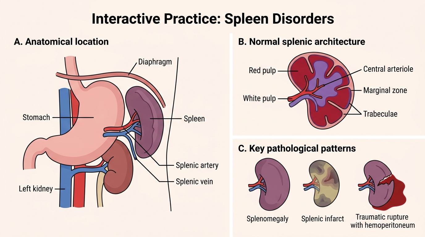
⚑ AI image — pending faculty review (auto-QA score 6/10; best of 3 attempts)

A multi-panel medical education diagram reviews normal spleen anatomy and key spleen disorders including splenomegaly, hypersplenism, infarction, and rupture.

Spleen Disorders: Interactive Review Map

Panel A: Normal spleen anatomy showing spleen, ribs 9-11, stomach, left kidney, diaphragm, splenic artery, and splenic vein. Panel B: Congestive splenomegaly showing enlarged spleen, capsule, red pulp expansion, and increased blood pooling. Panel C: Hypersplenism schematic showing enlarged spleen causing reduced RBCs, WBCs, and platelets. Panel D: Splenic infarct showing splenic arterial branch occlusion and wedge-shaped pale infarct. Panel E: Splenic rupture showing capsular tear, splenic laceration, and intraperitoneal bleeding. Footer: Interactive practice cues showing crossword for key terms and flashcards for disorder recognition.

Reinforce the key terms and concepts from this module with these self-check activities.

Interactive practice: Crossword

Interactive practice: Flashcards