Page 1 of 4
AN32.1-2 | Anterior Triangle — SDL Guide
Learning Objectives
- Describe the boundaries and subdivisions of the anterior triangle of the neck (AN32.1)
- Describe and demonstrate the boundaries and contents of the muscular, carotid, digastric, and submental triangles (AN32.2)
INSTRUCTIONS
Work systematically through each sub-triangle. Use palpation landmarks (hyoid, thyroid cartilage, sternocleidomastoid) to anchor the anatomy.
References
- Gray's Anatomy for Students — Head and Neck: Anterior Triangle (Textbook)
- BD Chaurasia's Human Anatomy Vol. 3 — Neck (Textbook)
- Last's Anatomy — Neck and Head (Textbook)
Version 1.0 | Academe Content Engine v2, MGMCRI Department of Anatomy
CLINICAL SCENARIO
A 40-year-old woman presents to a government hospital in Chennai with a painless swelling in the anterior aspect of the neck that moves on swallowing and on protrusion of the tongue. On examination, it appears to lie in the midline, below the hyoid bone, and feels cystic.
What structure has given rise to this swelling? In which sub-triangle of the anterior neck does it lie? Why does it move with both swallowing and tongue protrusion?
By the end of this module, you will have mastered the anatomy that explains this presentation — a thyroglossal cyst.
WHY THIS MATTERS
The anterior triangle of the neck is one of the most clinically important regions in surgery. As a doctor in India, you will encounter:
- Thyroglossal cysts and fistulae — midline swellings; surgically removed via Sistrunk's operation (must remove hyoid body to prevent recurrence)
- Carotid body tumour (chemodectoma) — at the carotid bifurcation; pulsatile, transmits pulsation, split sign on carotid angiography
- Cervical lymphadenopathy — tuberculous lymphadenitis is the most common cause of neck swelling in India; lies in the carotid triangle
- Carotid endarterectomy — surgical treatment for carotid artery stenosis; detailed knowledge of carotid triangle anatomy is essential
- Deep space neck infections — retropharyngeal and parapharyngeal abscesses; understanding the fascial planes of the neck prevents misdiagnosis
RECALL
Before we begin, recall the boundaries of the neck triangles:
- The sternocleidomastoid (SCM) divides the neck into anterior and posterior triangles
- The anterior triangle: bounded anteriorly by the midline of the neck, posteriorly by SCM, superiorly by the inferior border of the mandible
- The hyoid bone lies at the C3 vertebral level; it is the only bone in the body with no bony articulation
- The thyroid cartilage (Adam's apple) lies at C4–C5 level; the cricoid cartilage lies at C6
Anterior Triangle — Boundaries and Subdivisions (AN32.1)
Overall Boundaries of the Anterior Triangle:
- Anterior (medial) boundary: Midline of neck (from chin to sternum)
- Posterior (lateral) boundary: Anterior border of sternocleidomastoid muscle
- Superior (upper) boundary: Lower border of the mandible + a line continued to the mastoid process
- Floor: Pharynx, larynx, thyroid gland
- Roof: Investing layer of deep cervical fascia + platysma
Subdivision by the Digastric and Omohyoid Muscles:
The anterior triangle is divided into four sub-triangles by the:
1. Anterior belly of digastric (superomedial)
2. Posterior belly of digastric (superolateral)
3. Superior belly of omohyoid (inferolateral)
The four sub-triangles are:
| Sub-triangle | Boundaries | Key Contents |
|---|---|---|
| Submental | Anterior bellies of digastric (both sides) + hyoid | Submental lymph nodes, small veins |
| Digastric (Submandibular) | Mandible (above) + both bellies of digastric (below) | Submandibular gland + lymph nodes, facial artery + vein, hypoglossal nerve (CN XII), lingual nerve |
| Carotid | SCM (posterolateral) + posterior belly of digastric (above) + superior belly of omohyoid (below) | Common/internal/external carotid artery, internal jugular vein, CN IX, X, XI, XII, ansa cervicalis |
| Muscular | SCM (posterolateral) + superior belly of omohyoid (above) + midline (medially) | Infrahyoid (strap) muscles, thyroid and parathyroid glands, trachea, oesophagus |

Figure: Anterior Triangle — Boundaries and Subdivisions (AN32.1)
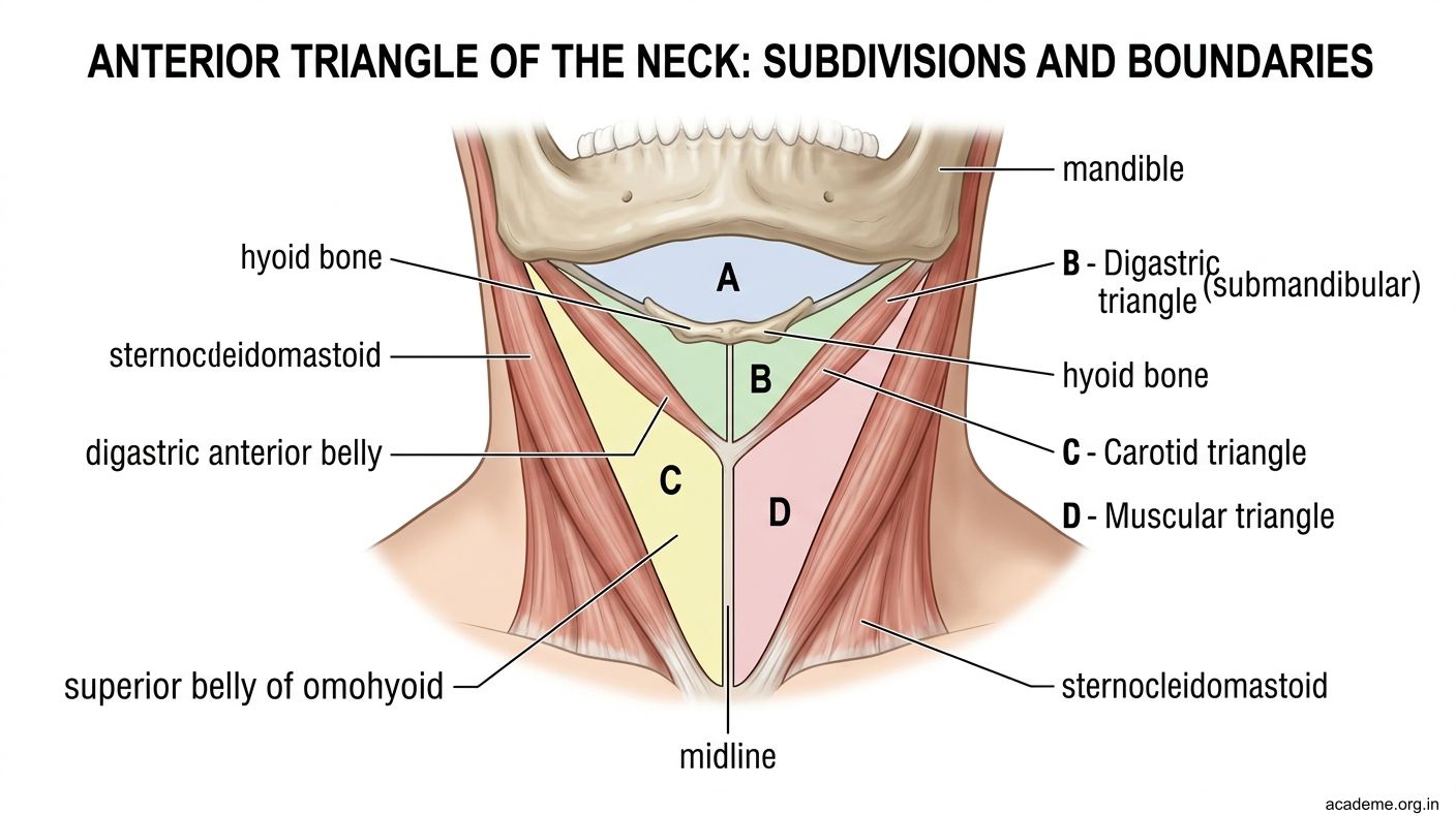
Figure: Anterior Triangle — Boundaries and Subdivisions (AN32.1)

Figure: Subdivision by the Digastric and Omohyoid Muscles:
Contents of Each Sub-Triangle (AN32.2)
1. Submental Triangle
- Boundaries: Anterior bellies of both digastric muscles laterally; hyoid bone inferiorly; midline of chin–hyoid superiorly
- Contents: Submental lymph nodes (drain the tip of tongue, floor of mouth, incisor teeth, central lower lip, chin skin); minute submental veins
- Clinical: Submental lymph nodes are enlarged in intra-oral infections and carcinoma of the floor of mouth
2. Digastric (Submandibular) Triangle
- Boundaries: Mandible (superiorly); anterior belly of digastric (anteromedially); posterior belly of digastric + stylohyoid (posterolaterally)
- Floor: Mylohyoid (anterior part), hyoglossus + part of middle pharyngeal constrictor (posterior part)
- Roof: Investing fascia + platysma + skin
Important contents:
• Submandibular gland — superficial part lies in the triangle; deep part extends around the posterior border of mylohyoid into the floor of the mouth. Wharton's duct opens beside the frenulum of the tongue.
• Facial artery — enters the triangle by hooking under the posterior belly of digastric; grooves the posterior surface of the submandibular gland; crosses the inferior border of mandible at the anterior edge of masseter (palpable pulse); continues on the face
• Facial vein — runs superficial and posterior to the artery; joins the anterior division of the retromandibular vein to form the common facial vein → drains into the internal jugular vein
• Submandibular lymph nodes — 3–6 nodes; drain the tongue (lateral 2/3), floor of mouth, cheek, lip; important in head and neck cancer staging
• Hypoglossal nerve (CN XII) — runs on the hyoglossus; supplies all intrinsic + extrinsic tongue muscles (except palatoglossus)
• Mylohyoid nerve (branch of inferior alveolar nerve) — runs in the groove on mylohyoid; sensory to the floor of the mouth
3. Carotid Triangle
- Boundaries: SCM (posterolaterally); posterior belly of digastric + stylohyoid (superiorly); superior belly of omohyoid (inferiorly)
- Floor: Thyrohyoid, hyoglossus, inferior and middle pharyngeal constrictors, longus colli
Important contents:
• Common carotid artery → bifurcates at the upper border of the thyroid cartilage (C4 level) into:
- Internal carotid artery (ICA) — no branches in the neck; runs posterolateral; enters carotid canal
- External carotid artery (ECA) — multiple branches (superior thyroid, lingual, facial, ascending pharyngeal, occipital, posterior auricular, and terminates as maxillary + superficial temporal)
• Carotid sinus — slight dilation at the ICA origin; contains baroreceptors (CN IX — Hering's nerve); stimulation causes reflex bradycardia + hypotension
• Carotid body — small chemoreceptor organ at the carotid bifurcation; responds to hypoxia, hypercapnia, pH changes; innervated by CN IX
• Internal jugular vein (IJV) — lateral to the carotid; joins the subclavian vein to form the brachiocephalic vein behind the sternoclavicular joint
• Vagus nerve (CN X) — between the carotid artery and IJV within the carotid sheath
• Hypoglossal nerve (CN XII) — curves forward across the carotid arteries (external, then internal); gives ansa cervicalis descendens before passing to the tongue
• Ansa cervicalis — loop on the anterior surface of the IJV; C1 fibres (superior root) + C2–C3 fibres (inferior root); innervates all infrahyoid (strap) muscles except thyrohyoid (directly by C1 via CN XII)
4. Muscular Triangle
- Boundaries: SCM (posterolaterally); superior belly of omohyoid (superolaterally); midline (medially)
- Contents:
- Infrahyoid (strap) muscles: sternohyoid, sternothyroid, thyrohyoid, omohyoid (superior belly)
- Thyroid gland — highly vascular; isthmus crosses the 2nd–4th tracheal rings; two lateral lobes; blood supply from superior thyroid (ECA) and inferior thyroid (thyrocervical trunk of subclavian) arteries
- Parathyroid glands (4 in total) — posterior surface of thyroid; superior pair at C5–C6 level; inferior pair variable (may be in thymus)
- Trachea — midline; thyroidectomy risk: recurrent laryngeal nerve injury causing hoarseness
- Oesophagus — posterior to trachea

Figure: Contents of Each Sub-Triangle (AN32.2)

Figure: Anterior Triangle — Key Points
SELF-CHECK — : Anterior Triangle
A midline neck swelling below the hyoid bone that moves with swallowing AND tongue protrusion is most likely:
A. Branchial cyst
B. Thyroglossal cyst
C. Lymph node
D. Lipoma
Reveal Answer
Answer: B. Thyroglossal cyst
The carotid bifurcation — where the common carotid divides into internal and external carotid arteries — lies at which vertebral level?
A. C2
B. C3
C. C4 (upper border of thyroid cartilage)
D. C6 (cricoid cartilage level)
Reveal Answer
Answer: C. C4 (upper border of thyroid cartilage)
The ansa cervicalis innervates all infrahyoid muscles EXCEPT which one (supplied directly by C1 via CN XII)?
A. Sternohyoid
B. Omohyoid
C. Thyrohyoid
D. Sternothyroid
Reveal Answer
Answer: C. Thyrohyoid
CLINICAL PEARL
Thyroglossal Cyst — Why Sistrunk's Operation Removes the Hyoid
The thyroglossal duct descends from the foramen cecum of the tongue (at the junction of the anterior 2/3 and posterior 1/3) through the midline, passing through (or around) the body of the hyoid bone, to reach the final position of the thyroid gland. A remnant of this tract that cystifies forms a thyroglossal cyst.
The duct invariably passes through the body of the hyoid bone in most people. If only the cyst is excised without removing the hyoid body and the suprahyoid tract segment, the cyst recurs (residual duct epithelium). Sistrunk's operation (excision of the cyst + central portion of the hyoid body + the tract up to the foramen cecum) achieves a cure rate of >95%.
This is why the cyst moves on tongue protrusion — the remaining attachment to the foramen cecum pulls it upward.
REFLECT
A patient in the carotid triangle has a pulsatile, mobile swelling that can be moved side-to-side but not up-and-down (the "split sign" on angiography). What is the likely diagnosis? Which cranial nerves are at risk during surgical removal, and why?
KEY TAKEAWAYS
Anterior Triangle — Key Points
- Boundaries: Midline (medially), SCM (laterally), mandible (superiorly).
- Sub-triangles: Submental, Digastric (submandibular), Carotid, Muscular.
- Carotid triangle: Most surgically important; contains carotid bifurcation at C4, IJV, CN IX/X/XI/XII, ansa cervicalis.
- Digastric triangle: Submandibular gland + lymph nodes, facial artery + vein, CN XII.
- Submental triangle: Only midline triangle; submental lymph nodes.
- Muscular triangle: Thyroid gland, strap muscles, trachea.
- Carotid sinus (baroreceptor, CN IX) + carotid body (chemoreceptor) at bifurcation.
- Ansa cervicalis (C1–C3): innervates infrahyoid muscles; thyrohyoid separately by C1 via CN XII.