Page 2 of 6
AN62.1-6 | Cranial nerve nuclei & Cerebral hemispheres — SDL Guide (Part 2)
Part 4: Basal Ganglia & Limbic Lobe (AN62.4)
Basal Ganglia Components:
• Striatum = Caudate nucleus + Putamen (connected anteriorly)
• Lenticular nucleus = Putamen + Globus pallidus (external GPe + internal GPi)
• Corpus striatum = Striatum + Lenticular nucleus
• Basal ganglia (functional group) = Corpus striatum + Subthalamic nucleus + Substantia nigra
Direct and Indirect Pathways:
Direct pathway (facilitatory — promotes movement):
Cortex → Striatum → GPi (inhibited) → Thalamus disinhibited → Motor cortex activated → MOVEMENT
Indirect pathway (inhibitory — suppresses movement):
Cortex → Striatum → GPe (inhibited) → Subthalamic nucleus (disinhibited) → GPi (activated) → Thalamus inhibited → Motor cortex suppressed → NO MOVEMENT
Balance between direct and indirect = normal smooth movement
Pathophysiology of Movement Disorders:
| Disorder | Pathology | Dominant Pathway | Clinical |
|---|---|---|---|
| Parkinson's disease | Loss of SN dopamine → reduces direct pathway, enhances indirect | Indirect dominant | Bradykinesia, rigidity, resting tremor |
| Huntington's disease | Loss of striatal neurons (indirect pathway first) | Direct dominant | Chorea (choreiform movements) |
| Hemiballismus | Damage to subthalamic nucleus (Patient C) | Indirect pathway loses STN inhibition | Violent flinging movements of contralateral limb |
| Athetosis | Basal ganglia lesion (often perinatal) | — | Slow writhing movements |
Limbic Lobe:
• Components: Cingulate gyrus, Parahippocampal gyrus, Hippocampus + dentate gyrus, Amygdala, Fornix → mammillary bodies → thalamus (anterior) → cingulate gyrus (Papez circuit)
• Hippocampus — memory consolidation (HM patient landmark); bilateral hippocampal damage → anterograde amnesia
• Amygdala — fear, emotion, aggression; lesion → Klüver-Bucy syndrome (placidity, hypersexuality, oral exploration)
• Fornix — main output of hippocampus → mammillary bodies → via mammillothalamic tract → anterior thalamus → cingulate (Papez circuit)
• TLE (temporal lobe epilepsy) = most common focal epilepsy in India; hippocampal sclerosis; can present with déjà vu, automatisms, fear
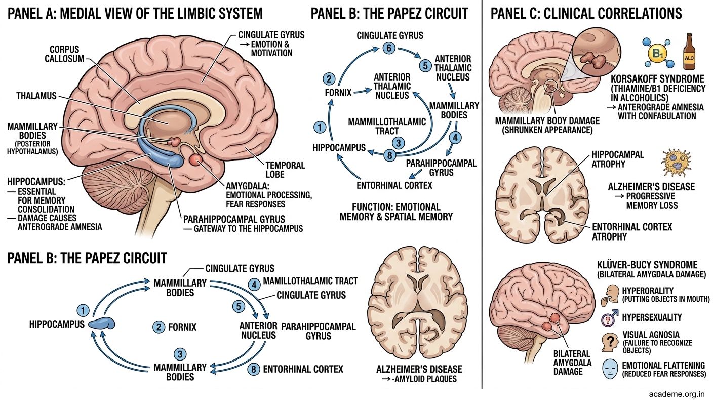
Figure: Direct and Indirect Pathways:
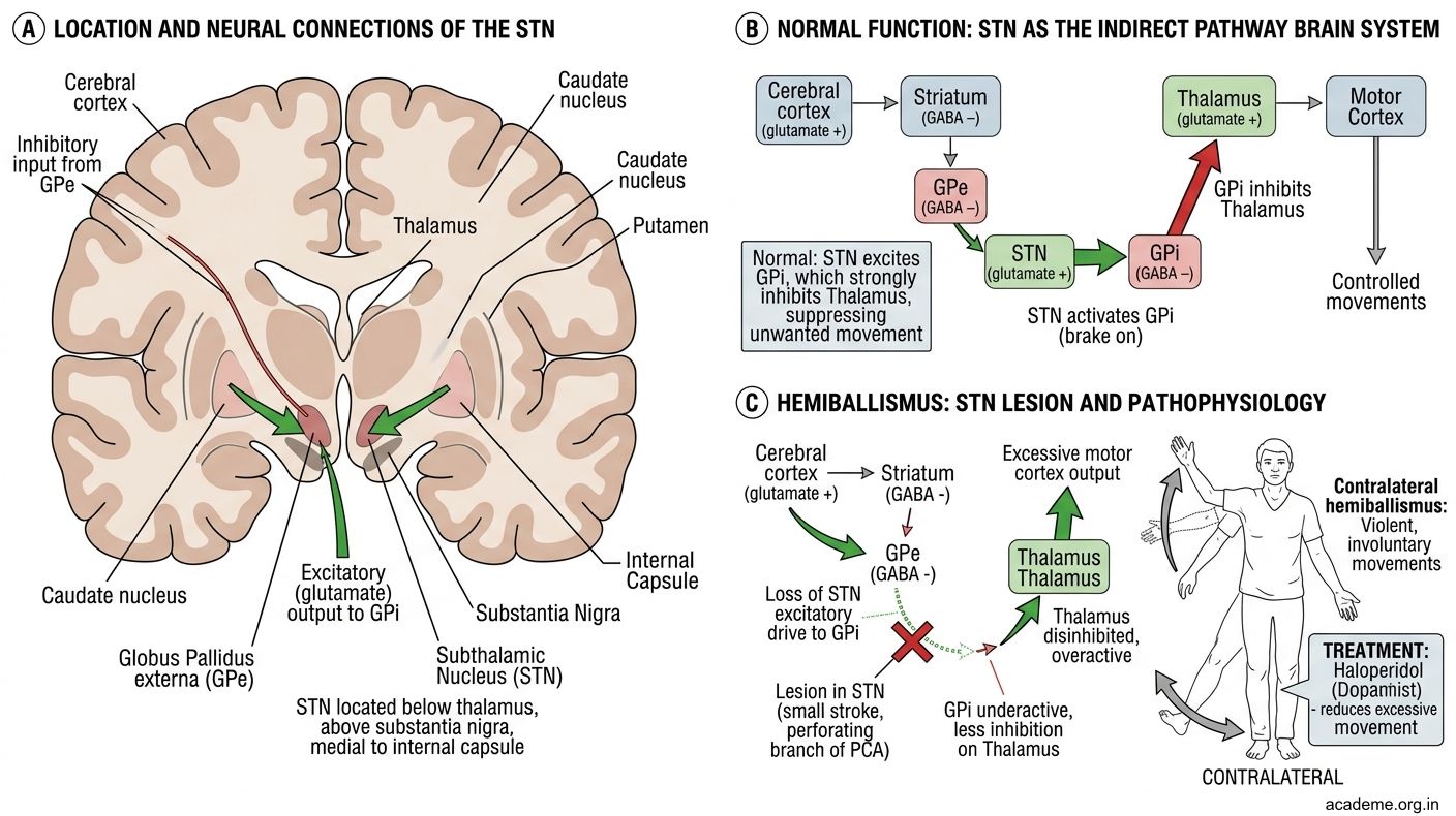
Figure: Pathophysiology of Movement Disorders:
Part 5: Thalamus, Hypothalamus & Diencephalon (AN62.5)
Thalamus (Dorsal Thalamus):
• Egg-shaped, bilateral, flanking the 3rd ventricle
• "Gateway to consciousness" — all sensory modalities (except smell) relay here before cortex
• Key nuclei:
| Nucleus | Input | Output | Function |
|---|---|---|---|
| VPL (ventroposterolateral) | Medial lemniscus, spinothalamic (body) | Somatosensory cortex (areas 1,2,3) | Body sensation |
| VPM (ventroposteromedial) | Trigeminal lemniscus (face) | Somatosensory cortex | Face sensation |
| VL (ventrolateral) | Cerebellum (dentate), basal ganglia | Motor cortex (area 4,6) | Motor coordination |
| LGN (lateral geniculate) | Optic tract (visual) | Primary visual cortex (area 17) | Vision |
| MGN (medial geniculate) | Lateral lemniscus (auditory) | Primary auditory cortex | Hearing |
| Anterior | Mammillothalamic tract | Cingulate gyrus | Limbic (Papez circuit) |
| CM (centromedian) | Basal ganglia, reticular formation | Striatum, cortex | Consciousness, arousal |
Hypothalamus:
• Floor and lower walls of 3rd ventricle; above pituitary stalk
• Controls: pituitary, autonomic NS, temperature, hunger/satiety, water balance (ADH), circadian rhythm, sleep-wake
• Key nuclei:
- Supraoptic (SON) and Paraventricular (PVN) → synthesise ADH (vasopressin) and oxytocin → released from posterior pituitary
- Anterior → parasympathetic, heat dissipation; Posterior → sympathetic, heat conservation
- Lateral → hunger centre (destroyed → starvation); Ventromedial → satiety centre (destroyed → hyperphagia)
Epithalamus: Pineal gland (melatonin, circadian rhythm) + habenular nuclei
Metathalamus: LGN + MGN (sensory relay for vision and hearing)
Subthalamus: Subthalamic nucleus (STN) — part of basal ganglia indirect pathway; damage → hemiballismus (Patient C in hook)
Part 6: Circle of Willis (AN62.6)
Formation:
• Formed by anastomosis of the anterior and posterior circulations at the base of the brain in the interpeduncular cistern

Figure: Part 6: Circle of Willis (AN62.6)
Vessels forming the circle (anterior → posterior, each side):
1. Anterior cerebral artery (ACA) — from ICA; supplies medial surface of hemisphere (motor/sensory for leg)
2. Anterior communicating artery (AComm) — connects left and right ACA; most common site of berry aneurysm
3. Middle cerebral artery (MCA) — largest branch of ICA; lateral surface (arm, face, Broca, Wernicke); most common stroke territory in India
4. Posterior communicating artery (PComm) — connects ICA to PCA; 2nd most common aneurysm site; compresses CN III (oculomotor)
5. Posterior cerebral artery (PCA) — from basilar artery; occipital lobe (visual cortex), thalamus, medial temporal
6. Basilar artery — from union of two vertebral arteries; gives anterior inferior cerebellar (AICA), posterior inferior cerebellar (PICA via vertebral), superior cerebellar (SCA)
Common aneurysm sites (berry aneurysms = congenital, at arterial bifurcations):
| Site | % |
|---|---|
| Anterior communicating artery | 30–35% |
| Posterior communicating artery | 25–30% |
| MCA bifurcation | 20% |
| Basilar tip | 5% |
| Others | <10% |
Circle of Willis Strokes:
• ACA territory: Leg weakness + sensory loss (contralateral) + frontal lobe signs; abulia, urinary incontinence (paracentral lobule)
• MCA territory: Arm + face weakness + sensory loss + aphasia (dominant hemisphere); contralateral homonymous hemianopia
• PCA territory: Contralateral homonymous hemianopia (occipital cortex) + thalamic pain syndrome; memory impairment (temporal);
• Lacunar infarcts — small perforating arteries (lenticulostriate from MCA, thalamoperforators from PCA) → pure motor or sensory strokes; most common in Indian hypertensives
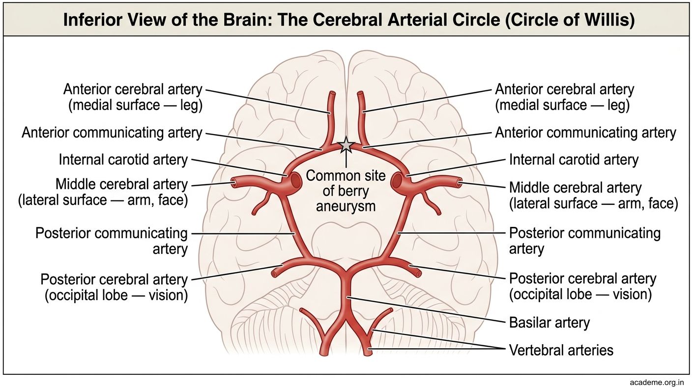
Figure: Part 6: Circle of Willis (AN62.6)
SELF-CHECK — : Cerebral Hemispheres
Broca's aphasia (motor aphasia) results from damage to areas 44 and 45 in the left hemisphere. The patient's speech is:
A. Non-fluent (effortful, halting) but comprehension is relatively preserved
B. Fluent but incomprehensible (jargon, paraphasias)
C. Complete absence of speech with normal comprehension
D. Preserved speech but inability to repeat
Reveal Answer
Answer: A. Non-fluent (effortful, halting) but comprehension is relatively preserved
Hemiballismus (violent flinging movements of one side of the body) results from damage to:
A. Caudate nucleus
B. Globus pallidus
C. Subthalamic nucleus (contralateral)
D. Putamen
Reveal Answer
Answer: C. Subthalamic nucleus (contralateral)
A pure motor stroke affecting the face, arm, and leg on one side with no sensory loss is most likely a lacunar infarct in the:
A. Posterior limb of the internal capsule and VPL thalamus
B. Posterior limb of the internal capsule (corticospinal and corticobulbar fibres)
C. Anterior limb of the internal capsule
D. Genu of the internal capsule alone
Reveal Answer
Answer: B. Posterior limb of the internal capsule (corticospinal and corticobulbar fibres)
CLINICAL PEARL
MCA Stroke — Most Common Stroke in India
Left MCA territory (dominant hemisphere) infarct causes the classic triad:
1. Right hemiplegia (arm > leg, face included — lateral motor cortex + internal capsule)
2. Broca's (expressive) aphasia or Wernicke's (receptive) aphasia depending on sub-territory
3. Right homonymous hemianopia (optic radiation involvement)
CT brain in acute MCA stroke:
• Early signs (<6h): hyperdense MCA sign (clot in vessel), loss of grey-white differentiation, sulcal effacement
• 24–72h: low-density wedge-shaped infarct in MCA territory
Indian thrombolysis reality: IV tPA (alteplase) within 4.5 hours is standard of care; mechanical thrombectomy (clot retrieval) within 24 hours for large vessel occlusion. Only 2–3% of Indian stroke patients reach hospital in time for thrombolysis — a major healthcare access challenge.