Page 1 of 4
BI1.1 | Basic Biochemistry — SDL Guide
This Self-Directed Learning guide introduces the molecular architecture of the living cell — the fundamental unit of life. By the end of this guide, you will be able to describe how a cell is organised at the molecular level, identify its sub-cellular components, and explain the composition and key functions of biological membranes.
Learning Objectives
- Describe the molecular and functional organisation of a typical cell (BI1.1)
- Identify the major sub-cellular components (organelles) and their functions
- Explain the composition of the biological membrane (fluid mosaic model)
- Describe the key functions of biological membranes including selective transport, communication, and compartmentalisation
INSTRUCTIONS
Read each section in order. Bold terms (like this) are key vocabulary — each is defined the first time it appears. Attempt the self-check questions before revealing the answer. Complete the reflection at the end.
References
- OpenStax Anatomy and Physiology 2e, Chapters 2–3 (CC BY 4.0). openstax.org
- Stryer L et al. Biochemistry, 8th ed. W.H. Freeman.
- NMC UG CBUC 2024, Competency BI1.1
CLINICAL SCENARIO
Imagine you are the intern on call at 2 AM. A patient with diabetic ketoacidosis comes in — her cells are starving even though her blood is full of glucose. The cell membrane is refusing to let glucose in without insulin. Meanwhile, in the patient beside her, a man with a genetic enzyme defect cannot break down glycogen inside his lysosomes — those tiny sacs within his cells are swelling and destroying liver tissue.
Both emergencies trace back to the same root: what happens inside and at the boundary of a cell. Before you can understand disease at the molecular level, you need to understand the cell not as a diagram you once memorised, but as a precision machine operating at the nanometre scale.
That is where this guide begins.
WHY THIS MATTERS
Every disease you will study in the next five years has a cellular and molecular explanation:
• Drug action — almost all drugs work by crossing or binding to the cell membrane
• Genetic disease — mutations alter proteins made by organelles called ribosomes
• Cancer — uncontrolled division begins with signals sent across the cell membrane
• Nutritional disease — vitamins and minerals are co-factors for reactions happening in specific organelles
The NMC requires you to "describe the molecular and functional organisation of a cell" (BI1.1) because this is the shared language of all of medicine. Pathology, Pharmacology, Physiology — they all speak cell biology.
RECALL
From your NCERT Class 11 Biology, you already know:
• A cell has a nucleus (control centre), cytoplasm (gel-like interior), and a cell membrane (outer boundary)
• Key organelles: mitochondria (energy), ribosomes (protein synthesis), endoplasmic reticulum (processing), Golgi apparatus (packaging)
• The cell membrane is made of a phospholipid bilayer
• Cells perform metabolism — breaking down and building up molecules
In this guide, we move from that overview to the molecular level — what these structures are actually made of, how they are organised, and why that organisation enables life.
The Cell: A Precision Molecular Machine
Think of a cell like a modern pharmaceutical factory. Every factory has:
• A management office (nucleus) — holds the blueprints (DNA), issues instructions (mRNA)
• Assembly lines (ribosomes) — read the blueprints and manufacture products (proteins)
• Processing and dispatch (endoplasmic reticulum + Golgi) — refine and package products
• Power plant (mitochondria) — generates energy to run all operations
• Waste disposal (lysosomes) — break down old parts and foreign materials
• Security fence (cell membrane) — controls what enters and exits
This is the concept of molecular organisation — the idea that a cell's physical structure directly determines its biological function. Nothing inside a cell is random; every molecule is positioned to do a specific job.
The basic unit of this organisation is the biomolecule — a carbon-containing molecule produced by living cells. The four classes of biomolecules are:
1. Carbohydrates — energy storage and cell recognition (e.g., glycogen, glucose)
2. Lipids — membrane structure, energy storage, and signalling (e.g., phospholipids, cholesterol)
3. Proteins — structural scaffolding, enzymes, transporters, receptors (most versatile)
4. Nucleic acids — information storage (DNA) and information transfer (RNA)
Every organelle is built from these four classes — and every disease involves at least one of them malfunctioning.
Etymology note: Biomolecule (from Greek bios = life + Latin molecula = small mass). A molecule is a biomolecule only if it is produced by, or essential to, living organisms.
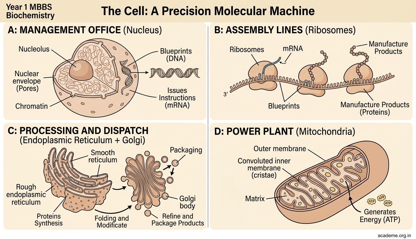

Figure: The Cell: A Precision Molecular Machine
Sub-cellular Components: The Organelles
The cell's interior is not a bag of soup. It is a highly organised collection of organelles (from Latin organella = little organ) — membrane-bound compartments, each with a specific molecular composition and function.
The major organelles and what they do:
| Organelle | Membrane? | Key Function |
|---|---|---|
| Nucleus | Double membrane (nuclear envelope) | Stores DNA; transcription of mRNA |
| Mitochondria | Double membrane | ATP synthesis (oxidative phosphorylation) |
| Rough ER | Single membrane + ribosomes | Synthesis and folding of secretory proteins |
| Smooth ER | Single membrane | Lipid synthesis; drug detoxification |
| Golgi apparatus | Single membrane | Modifies, sorts, and packages proteins/lipids |
| Lysosomes | Single membrane | Digestion of worn-out organelles and foreign material |
| Peroxisomes | Single membrane | Oxidative reactions; fatty acid breakdown |
| Ribosomes | No membrane | Protein synthesis (present in cytosol and on rough ER) |
| Cytoskeleton | Not an organelle | Provides shape, enables movement and division |
Important principle: The more metabolically active a cell, the more mitochondria it has. Cardiac muscle cells have ~5,000 mitochondria per cell. Mature red blood cells have none (they have no nucleus either — maximising space for haemoglobin).
Mnemonic for organelles: MERGE LPG
• Mitochondria — energy
• Endoplasmic reticulum — synthesis
• Ribosomes — protein factory
• Golgi — processing/packaging
• Endosomes/Lysosomes — digestion
• Lipid droplets — storage
• Peroxisomes — oxidation
• Glory (nucleus) — master control

Figure: Sub-cellular Components: The Organelles

Figure: The major organelles and what they do:
SELF-CHECK — 1 : Cell Organisation
A 28-year-old man presents with muscle weakness. Biopsy shows large, abnormal mitochondria and "ragged red fibres." Which cellular function is primarily affected?
A. Protein synthesis at the ribosomes
B. ATP generation through oxidative phosphorylation
C. Lipid packaging at the Golgi apparatus
D. DNA replication in the nucleus
Reveal Answer
Answer: B. ATP generation through oxidative phosphorylation
Which of the following is the correct sequence for a secretory protein, from synthesis to export?
A. Ribosome → Smooth ER → Golgi → Secretory vesicle → Exocytosis
B. Ribosome → Rough ER → Golgi → Secretory vesicle → Exocytosis
C. Nucleus → Ribosome → Lysosome → Exocytosis
D. Ribosome → Golgi → Rough ER → Secretory vesicle → Exocytosis
Reveal Answer
Answer: B. Ribosome → Rough ER → Golgi → Secretory vesicle → Exocytosis
The Biological Membrane: Composition and Structure
The biological membrane (or plasma membrane) is a selectively permeable barrier that separates the cell's interior from the external environment. It is not a rigid wall — think of it as a dynamic, fluid mosaic that is constantly in motion.
The Fluid Mosaic Model (Singer & Nicolson, 1972):
The membrane is described as:
• Fluid — the phospholipid molecules can move laterally (sideways) within each layer
• Mosaic — proteins are embedded like tiles within the lipid "sea"
Composition of the biological membrane:
1. Phospholipids (the backbone):
A phospholipid has a dual nature — crucial for membrane formation:
• Hydrophilic head (water-loving) — contains phosphate + glycerol, faces outward into water
• Hydrophobic tail (water-fearing) — two fatty acid chains, faces inward away from water
This creates a phospholipid bilayer — two sheets of phospholipids with their tails facing each other and heads facing outward. The result is a self-sealing, stable boundary that keeps the aqueous interior separate from the aqueous exterior.
Think of it like a sandwich: the bread (hydrophilic heads) is visible on both sides, the filling (hydrophobic tails) is hidden in the middle.
2. Proteins (the workers):
Two types are embedded in the bilayer:
• Integral proteins — span the full width of the membrane (transmembrane proteins); form channels, pumps, and receptors
• Peripheral proteins — attached to the surface only; involved in signalling and structural support
3. Cholesterol (the stabiliser):
Scattered between phospholipid molecules, cholesterol acts as a "fluidity buffer":
• At high temperatures: stiffens the membrane (prevents it becoming too fluid)
• At low temperatures: prevents crystallisation (prevents it becoming too rigid)
• Result: membrane fluidity is maintained across a range of temperatures — essential for a warm-blooded mammal
4. Glycoproteins and Glycolipids (the ID badges):
Glycoproteins (protein + sugar chain) and glycolipids (lipid + sugar chain) project from the outer leaflet only. They form the glycocalyx — a fuzzy coat on the cell surface that functions in:
• Cell recognition (self vs. non-self — the basis of immunity and blood group typing)
• Cell-to-cell communication and adhesion
• Protection from mechanical damage


Figure: The Biological Membrane: Composition and Structure

Figure: Composition of the biological membrane:
Functions of the Biological Membrane
The membrane is not a passive barrier — it is an active, intelligent gatekeeper. Its functions fall into four categories:
1. Selective Permeability (transport)
The membrane controls what enters and exits by three mechanisms:
Passive transport (no energy required):
• Simple diffusion — small, non-polar molecules (O₂, CO₂, alcohol) dissolve through the lipid bilayer freely, following concentration gradients
• Osmosis — water moves through aquaporins (water channel proteins) from low solute → high solute concentration
• Facilitated diffusion — large or polar molecules (glucose, amino acids) cannot cross alone; they need channel proteins or carrier proteins
Active transport (energy required — ATP):
• The Na⁺/K⁺-ATPase pump (sodium-potassium pump) moves 3 Na⁺ OUT and 2 K⁺ IN against concentration gradients, consuming 1 ATP per cycle. This pump maintains the electrical charge difference across the membrane that allows nerve and muscle function.
• Clinically: cardiac glycosides (digoxin) work by inhibiting this pump in heart muscle.
Vesicular transport:
• Endocytosis — membrane wraps around material and brings it in:
- Phagocytosis: engulfs large particles (bacteria, dead cells) — your neutrophils do this
- Pinocytosis: takes up small droplets of fluid
- Receptor-mediated endocytosis: takes up specific molecules (LDL cholesterol, iron-transferrin)
• Exocytosis — vesicles fuse with membrane and release contents outside (insulin secretion, neurotransmitter release)
2. Cell Signalling and Reception
Membrane receptor proteins bind specific chemical signals (ligands) — hormones, neurotransmitters, growth factors. This binding triggers a cascade of events inside the cell without the signal itself entering. Example: insulin binds the insulin receptor on fat and muscle cells → glucose transporters (GLUT4) move to the surface → glucose enters.
3. Compartmentalisation
Internal membranes divide the cell into distinct compartments (the organelles). This allows:
• Incompatible reactions to occur simultaneously (acidic lysosomes at pH 4.5 alongside neutral cytoplasm at pH 7.2)
• Concentrating enzymes and substrates for efficiency
• Protecting DNA from reactive metabolites
4. Structural Support and Cell Shape
The cytoskeleton (actin filaments, microtubules, intermediate filaments) connects to membrane proteins, giving the cell shape and enabling movement (cell migration, phagocytosis, mitosis).
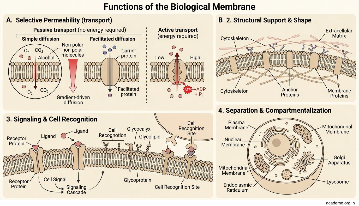
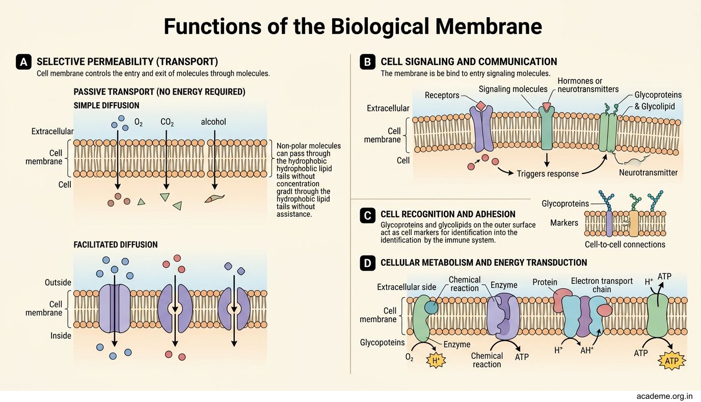

Figure: Functions of the Biological Membrane

Figure: Cystic Fibrosis — a membrane protein disease

Figure: Key Takeaways for BI1.1:
CLINICAL PEARL
Cystic Fibrosis — a membrane protein disease
Cystic fibrosis (CF) is caused by a mutation in the gene for CFTR (Cystic Fibrosis Transmembrane conductance Regulator) — an integral membrane protein that functions as a chloride ion channel. The most common mutation (ΔF508) causes the protein to misfold in the ER and never reach the membrane.
Without functional CFTR at the cell surface:
• Chloride cannot exit the cell → sodium (following electrical charge) stays in → water stays in → thick, sticky mucus forms
• Airways, pancreatic ducts, and sweat glands are all affected
The treatment revolution: drugs like ivacaftor (a potentiator) and lumacaftor (a corrector) work at the molecular level — one helps the mutant protein reach the membrane, the other helps it open properly. This is precision medicine at the membrane level.
In India, CF is rarer than in Europe, but the biochemical principle — that membrane protein dysfunction causes systemic disease — applies to many common Indian conditions: G6PD deficiency, hereditary spherocytosis, and many enzyme-linked receptor disorders.
SELF-CHECK — 2 : Biological Membrane
A patient's red blood cells lyse when placed in distilled water. Which membrane property explains this?
A. Selective permeability — water enters by osmosis until the cell bursts
B. Active transport — the Na+/K+ pump fails without energy
C. Receptor-mediated endocytosis — the cell engulfs excess water
D. Exocytosis — the cell releases internal contents into the water
Reveal Answer
Answer: A. Selective permeability — water enters by osmosis until the cell bursts
Which component of the biological membrane is primarily responsible for "cell recognition" and determines blood group type?
A. Phospholipid tails (hydrophobic core)
B. Cholesterol molecules (fluidity buffer)
C. Glycoproteins and glycolipids (glycocalyx)
D. Peripheral proteins (surface attachment)
Reveal Answer
Answer: C. Glycoproteins and glycolipids (glycocalyx)
REFLECT
Take 5 minutes to connect what you have learned to your own body:
- Palpate the back of your hand — the skin you see and feel is built from billions of cells, each surrounded by a phospholipid bilayer that is right now maintaining concentration gradients and responding to signals.
- Consider your last meal — glucose from your food needed to cross the intestinal cell membrane via GLUT2 (facilitated diffusion), then enter your muscle cells via GLUT4 (insulin-dependent). How many membranes did a single glucose molecule cross to reach your muscle?
- Think about the clinical cases in the hook — the diabetic patient and the enzyme defect patient. Now explain to yourself (in 2–3 sentences) how each condition links to what you have learned about cell organisation and membrane function.
Write your answers in your learning portfolio before moving on.
KEY TAKEAWAYS
Key Takeaways for BI1.1:
- A cell is a molecularly organised precision machine — every structure reflects a specific function. The four classes of biomolecules (carbohydrates, lipids, proteins, nucleic acids) build all cellular components.
2. The major organelles and their functions:
• Nucleus → DNA storage and gene expression
• Mitochondria → ATP synthesis (oxidative phosphorylation)
• Rough/Smooth ER → protein and lipid synthesis/processing
• Golgi → modification, sorting, and packaging
• Lysosomes → intracellular digestion (pH 4.5)
• Ribosomes → protein synthesis (no membrane)
3. The biological membrane consists of:
• Phospholipid bilayer (selective permeability framework)
• Integral and peripheral proteins (channels, pumps, receptors)
• Cholesterol (fluidity buffer)
• Glycoproteins/glycolipids (cell recognition — glycocalyx)
4. Membrane functions:
• Selective transport: simple diffusion / osmosis / facilitated diffusion / active transport (Na⁺/K⁺ pump) / endocytosis / exocytosis
• Cell signalling via membrane receptor proteins
• Compartmentalisation (enables incompatible reactions simultaneously)
• Structural support
- Clinical bridge: Nearly every drug, toxin, and genetic disease involves either a membrane protein or an organelle. Understanding cell organisation is the foundation of all molecular medicine.
Review checklist: Can you define every bold term in this guide without looking? If not, use the glossary below.