Page 1 of 7
BI4.1-8 | Chemistry and Metabolism of Lipids — SDL Guide
Learning Objectives
- Describe the classification of lipids — simple, compound, and derived lipids — and their biological roles (BI4.1)
- Describe the chemistry and functions of fatty acids — saturated, unsaturated, essential, and non-essential (BI4.2)
- Describe the oxidation of fatty acids — beta-oxidation, energetics, and regulation (BI4.3)
- Describe the synthesis of fatty acids (lipogenesis) — pathway, regulation, and hormonal control (BI4.4)
- Describe the metabolism of cholesterol — synthesis (HMG-CoA reductase pathway), transport, and excretion (BI4.5)
- Describe the chemistry and metabolism of phospholipids and glycolipids — structure, function, and clinical significance (BI4.6)
- Describe the structure, function, and metabolism of lipoproteins — chylomicrons, VLDL, LDL, HDL — and their role in atherosclerosis (BI4.7)
- Describe the biochemical basis of dyslipidaemias and the mechanism of action of lipid-lowering drugs including statins (BI4.8)
INSTRUCTIONS
This module covers the chemistry and metabolism of lipids — the molecules that store energy, build membranes, and (when they go wrong) cause the world's leading cause of death. We'll start with what lipids ARE, then follow them through breakdown (beta-oxidation) and synthesis (lipogenesis), before focusing on the clinically critical topic: cholesterol, lipoproteins, and how they cause atherosclerosis.
Parallel connections: In Anatomy (AN22), you're studying the coronary arteries — the very arteries that atherosclerosis blocks to cause myocardial infarction. The LDL cholesterol you learn about today is literally what accumulates in those arterial walls. In Physiology (PY5), you're studying cardiovascular physiology — blood pressure regulation fails when vessels harden from atherosclerotic plaque.
References
- Harper's Illustrated Biochemistry, 31st ed., Chapters 21-26: Lipid Metabolism (textbook)
- Lehninger Principles of Biochemistry, 8th ed., Chapters 17, 21: Fatty Acid Metabolism and Lipid Biosynthesis (textbook)
- Lippincott's Illustrated Reviews: Biochemistry, 8th ed., Chapters 15-18: Lipid Metabolism (textbook)
- OpenStax Anatomy and Physiology 2e, Chapter 24: Metabolism and Nutrition (CC BY 4.0) (textbook (CC BY 4.0))
Version 2.0 | NMC CBUC 2024, Adapted from Harper's Illustrated Biochemistry
CLINICAL SCENARIO
Heart disease is the number 1 killer in India — and it starts with cholesterol in your blood vessels. The LDL cholesterol you'll learn about today is literally called 'bad cholesterol' in every clinic. But here's what most people don't know: cholesterol is not a villain. Your body makes about 1 gram of it every single day because you NEED it — for cell membranes, for steroid hormones, for bile acids that digest the food you eat. The problem starts when there's too much of it in the wrong place. By the end of this module, you'll understand exactly how a simple molecule of fat in your dal or ghee gets transformed into energy, membranes, hormones — or deadly arterial plaque.
WHY THIS MATTERS
As a doctor in India, you will encounter dyslipidaemia every single day. India has one of the highest rates of premature coronary artery disease in the world — Indians develop heart attacks a decade earlier than Western populations. The lipid profile (total cholesterol, LDL, HDL, triglycerides) is one of the most commonly ordered blood tests in clinical practice. Understanding lipid biochemistry is not optional — it's the foundation for prescribing statins (the world's most prescribed drug class), counselling patients about diet, and understanding why a 42-year-old IT professional in Chennai can have a heart attack despite being a vegetarian.
RECALL
From your study of carbohydrate metabolism (BI3), you know that glucose is the body's primary fuel. You also know about acetyl-CoA — the 2-carbon unit that enters the TCA cycle to generate ATP. Keep acetyl-CoA in mind: it's the universal currency of metabolism. Fatty acids are broken down INTO acetyl-CoA (beta-oxidation), and excess acetyl-CoA is used to BUILD fatty acids (lipogenesis). The TCA cycle and electron transport chain you studied in BI3 are the final common pathway for energy generation from ALL fuels — carbohydrates, fats, and proteins.
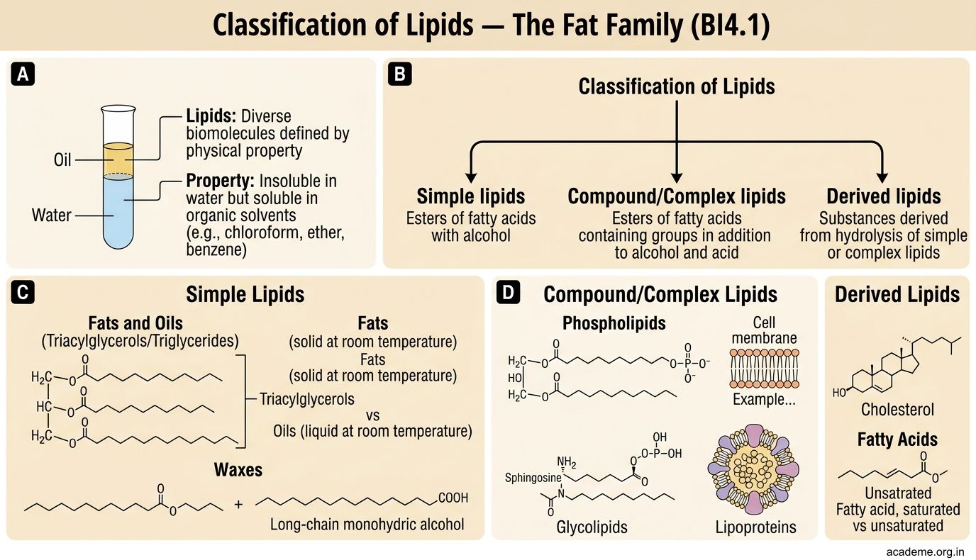
Classification of Lipids — The Fat Family (BI4.1)
Lipids are a diverse group of biomolecules defined by one property: they are insoluble in water but soluble in organic solvents (chloroform, ether, benzene). Unlike carbohydrates and proteins, lipids are not defined by a common structural backbone — they are defined by their physical behaviour.

Figure: Classification of Lipids — The Fat Family (BI4.1)
Lipids are classified into three main groups:
1. Simple lipids — esters of fatty acids with alcohols:
• Fats and oils (triacylglycerols/triglycerides) — three fatty acids esterified to glycerol. Fats are solid at room temperature (saturated, e.g. ghee, butter); oils are liquid (unsaturated, e.g. groundnut oil, olive oil). Triglycerides are the body's primary energy store — stored in adipose tissue, providing ~9 kcal/g (more than double the energy density of carbohydrates at ~4 kcal/g).
• Waxes — esters of fatty acids with long-chain alcohols. Found in sebum (skin), cerumen (earwax), and the coating of leaves.
2. Compound (complex) lipids — contain an additional group beyond fatty acids and alcohol:
• Phospholipids — contain a phosphate group. The most important membrane lipids. Two subtypes: glycerophospholipids (phosphatidylcholine, phosphatidylethanolamine — major components of cell membranes) and sphingophospholipids (sphingomyelin — abundant in the myelin sheath of nerves).
• Glycolipids — contain a sugar group. Cerebrosides (one sugar) and gangliosides (oligosaccharide with sialic acid) — abundant in brain and nervous tissue.
• Lipoproteins — lipids complexed with proteins for transport in blood (chylomicrons, VLDL, LDL, HDL — we'll cover these in Part 2).
3. Derived lipids — obtained by hydrolysis of simple or compound lipids:
• Fatty acids — the building blocks of most lipids
• Cholesterol — a sterol (steroid + alcohol). Precursor to bile acids, steroid hormones (cortisol, aldosterone, testosterone, oestrogen), and vitamin D.
• Prostaglandins, leukotrienes, thromboxanes — signalling molecules derived from arachidonic acid (a 20-carbon fatty acid). These mediate inflammation, pain, fever, and blood clotting.
Key concept: Lipids serve four major biological roles: (1) energy storage (triglycerides), (2) structural (phospholipids in membranes), (3) signalling (steroid hormones, prostaglandins), (4) insulation and protection (subcutaneous fat, myelin).
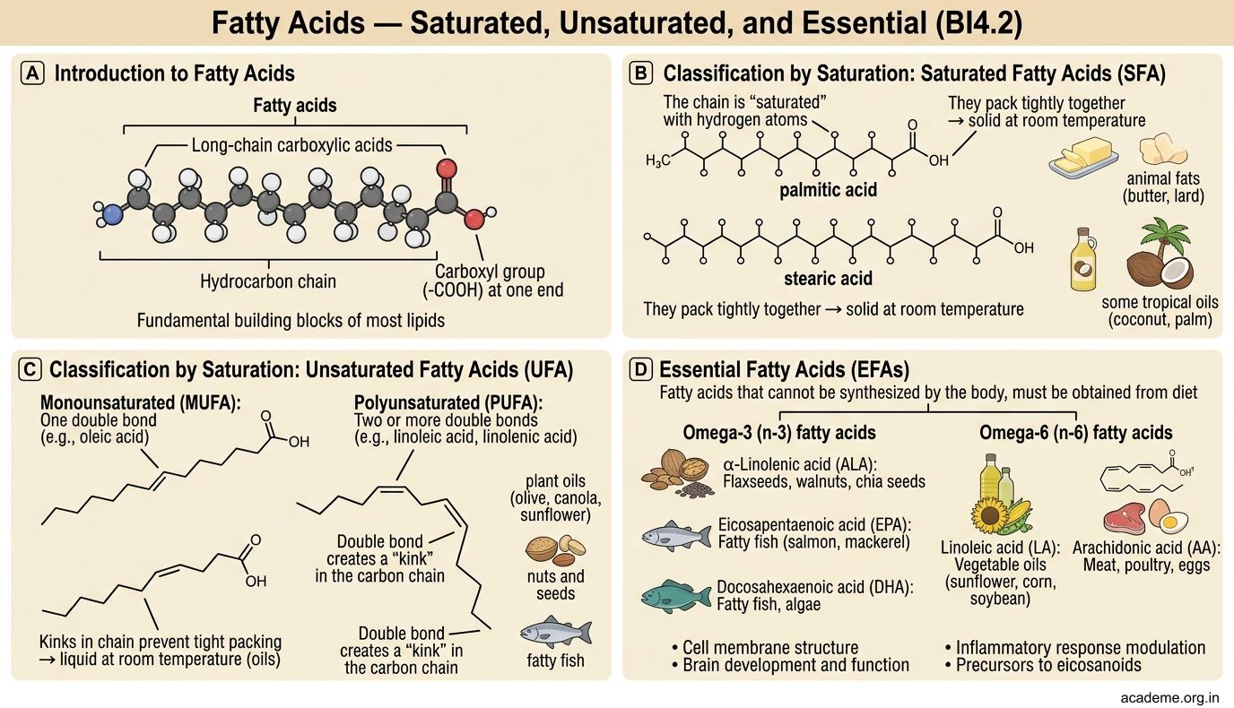
Fatty Acids — Saturated, Unsaturated, and Essential (BI4.2)
Fatty acids are long-chain carboxylic acids — a hydrocarbon chain with a carboxyl group (-COOH) at one end. They are the fundamental building blocks of most lipids.

Figure: Fatty Acids — Saturated, Unsaturated, and Essential (BI4.2)
Classification by saturation:
• Saturated fatty acids (SFA) — no double bonds in the carbon chain. The chain is 'saturated' with hydrogen atoms. They pack tightly together → solid at room temperature. Examples: palmitic acid (C16:0 — 16 carbons, 0 double bonds), stearic acid (C18:0). Found abundantly in ghee, butter, coconut oil.
- Monounsaturated fatty acids (MUFA) — one double bond (usually cis configuration, creating a 'kink' in the chain). Example: oleic acid (C18:1, Δ9 — 18 carbons, 1 double bond at position 9). Found in olive oil, groundnut oil, mustard oil. The kink prevents tight packing → liquid at room temperature.
- Polyunsaturated fatty acids (PUFA) — two or more double bonds. Examples: linoleic acid (C18:2, omega-6), linolenic acid (C18:3, omega-3), arachidonic acid (C20:4, omega-6). The more double bonds, the more fluid the molecule.
Essential fatty acids (EFAs):
Humans cannot synthesise double bonds beyond carbon 9 from the carboxyl end. Therefore, two fatty acids are essential (must come from diet):
• Linoleic acid (C18:2, omega-6) — found in sunflower oil, safflower oil, soybean oil
• α-Linolenic acid (C18:3, omega-3) — found in flaxseed, walnuts, fish oil
From linoleic acid, the body can synthesise arachidonic acid (the precursor to prostaglandins, thromboxanes, and leukotrienes). From α-linolenic acid, the body can (inefficiently) synthesise EPA and DHA — the omega-3 fatty acids in fish oil that are cardioprotective.
Omega numbering: The omega (ω) system counts from the methyl end (opposite to the carboxyl end). Omega-3 means the first double bond is 3 carbons from the methyl end. Omega-6 means 6 carbons from the methyl end. This is the system used in clinical nutrition.
Trans fatty acids: Industrial hydrogenation of vegetable oils creates trans double bonds (the hydrogen atoms are on opposite sides of the double bond, unlike the natural cis configuration). Trans fats behave like saturated fats — they raise LDL cholesterol and lower HDL cholesterol, increasing cardiovascular risk. The WHO has called for global elimination of industrial trans fats by 2023. Common sources in India: vanaspati ghee, bakery products, deep-fried street food.
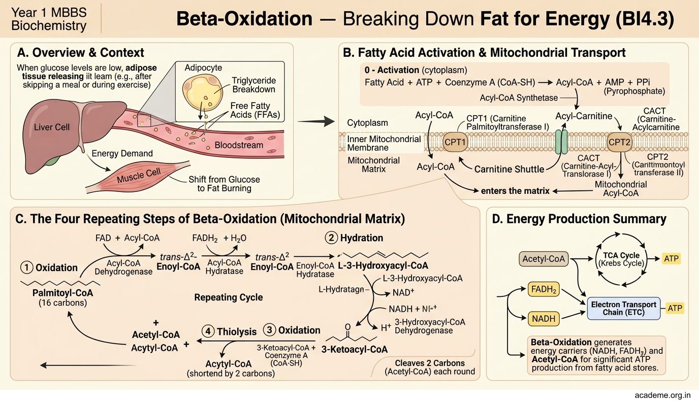
Beta-Oxidation — Breaking Down Fat for Energy (BI4.3)
When you skip a meal or exercise intensely, your body shifts from burning glucose to burning fat. Beta-oxidation is the process by which fatty acids are broken down into acetyl-CoA units, which then enter the TCA cycle for energy generation.

Figure: The 4-step spiral (each cycle removes 2 carbons):

Figure: Beta-Oxidation — Breaking Down Fat for Energy (BI4.3)
The process occurs in the mitochondrial matrix and has four repeating steps:
Step 0 — Activation (cytoplasm):
Before a fatty acid can enter the mitochondrion, it must be 'activated' by attaching to coenzyme A (CoA), forming acyl-CoA. This reaction requires 2 ATP (technically ATP → AMP + PPi, equivalent to 2 ATP) and is catalysed by acyl-CoA synthetase (also called fatty acid thiokinase).
The carnitine shuttle — getting the fatty acid across the mitochondrial membrane:
The inner mitochondrial membrane is impermeable to long-chain acyl-CoA. The fatty acid must be transferred to carnitine by CPT-I (carnitine palmitoyltransferase I, on the outer membrane), transported across as acylcarnitine by a translocase, and then transferred back to CoA by CPT-II (on the inner membrane). CPT-I is the rate-limiting step of beta-oxidation. It is inhibited by malonyl-CoA (the first committed intermediate of fatty acid synthesis) — this is how the body prevents simultaneous synthesis and breakdown of fatty acids.
The 4-step spiral (each cycle removes 2 carbons):
- Oxidation (by acyl-CoA dehydrogenase) — removes 2H → produces FADH₂
- Hydration (by enoyl-CoA hydratase) — adds H₂O across the double bond
- Oxidation (by 3-hydroxyacyl-CoA dehydrogenase) — removes 2H → produces NADH
- Thiolysis (by β-ketothiolase) — cleaves off acetyl-CoA + a shortened acyl-CoA
The shortened acyl-CoA re-enters the spiral, losing 2 carbons each cycle.
Energy yield from palmitic acid (C16:0):
• 7 cycles of beta-oxidation → 8 acetyl-CoA + 7 FADH₂ + 7 NADH
• 8 acetyl-CoA → TCA cycle → 8 × 10 ATP = 80 ATP
• 7 FADH₂ → 7 × 1.5 = 10.5 ATP
• 7 NADH → 7 × 2.5 = 17.5 ATP
• Total = 108 ATP − 2 ATP (activation) = 106 net ATP
Compare this to glucose: 1 glucose (6C) yields ~30-32 ATP. 1 palmitate (16C) yields 106 ATP. Fat is a far more efficient energy store — this is why the body stores excess energy as fat, not glycogen.
Clinical connection — MCAD deficiency:
Medium-chain acyl-CoA dehydrogenase (MCAD) deficiency is the most common inherited disorder of fatty acid oxidation. Affected children cannot oxidise medium-chain fatty acids → they develop hypoketotic hypoglycaemia during fasting (low blood sugar + no ketone bodies as backup). Newborn screening for MCAD deficiency is lifesaving — treatment is simply avoiding prolonged fasting.
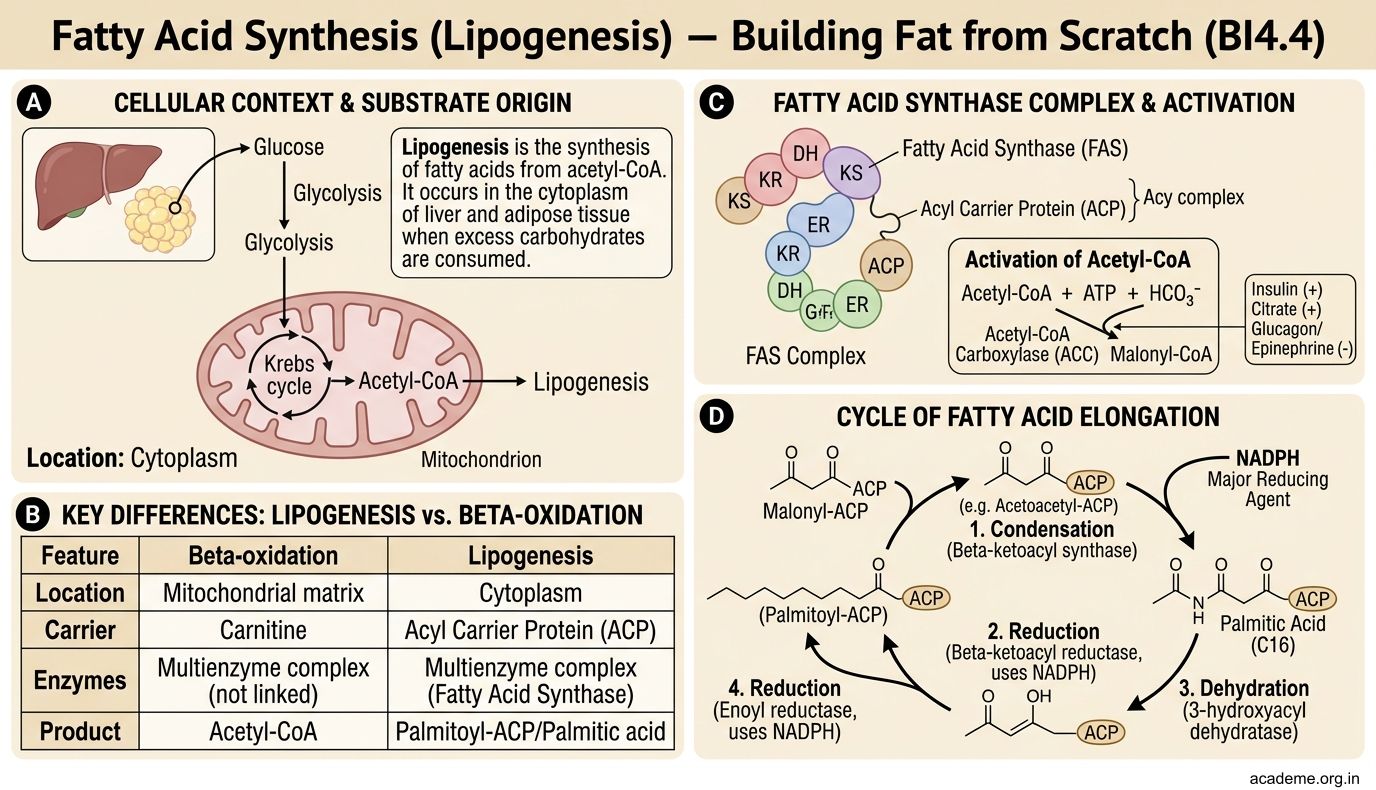
Fatty Acid Synthesis (Lipogenesis) — Building Fat from Scratch (BI4.4)
When you eat more carbohydrates than you need, your body converts the excess into fat. This is lipogenesis — the synthesis of fatty acids from acetyl-CoA. It occurs in the cytoplasm (not the mitochondrion) of liver and adipose tissue.

Figure: Key differences from beta-oxidation:

Figure: Fatty Acid Synthesis (Lipogenesis) — Building Fat from Scratch (BI4.4)
Key differences from beta-oxidation:
| Feature | Beta-oxidation | Lipogenesis |
|---|---|---|
| Location | Mitochondrial matrix | Cytoplasm |
| Carrier | CoA | ACP (acyl carrier protein) |
| 2-carbon unit | Acetyl-CoA (released) | Malonyl-CoA (added) |
| Coenzyme | FAD, NAD⁺ (reduced) | NADPH (oxidised) |
| Enzyme | Multiple separate enzymes | Fatty acid synthase (single multi-enzyme complex) |
| Regulation | Activated by fasting | Activated by feeding |
The pathway:
- Acetyl-CoA must exit the mitochondrion — it does so as citrate (via the citrate shuttle: acetyl-CoA + OAA → citrate in mitochondrion, citrate crosses membrane, ATP-citrate lyase regenerates acetyl-CoA + OAA in cytoplasm).
2. Committed step: Acetyl-CoA → malonyl-CoA by acetyl-CoA carboxylase (ACC) — this is the rate-limiting enzyme of fatty acid synthesis. It requires biotin as a cofactor and is:
- Activated by: citrate, insulin (fed state)
- Inhibited by: palmitoyl-CoA (product feedback), glucagon/adrenaline (fasting/stress)
- Fatty acid synthase (FAS) — a remarkable multi-enzyme complex that performs 7 sequential reactions, adding 2 carbons (from malonyl-CoA) per cycle. It uses NADPH as the reducing agent (supplied by the pentose phosphate pathway and the malic enzyme).
- The product is palmitic acid (C16:0) — further elongation and desaturation occur in the endoplasmic reticulum.
Hormonal regulation — the fed vs fasting switch:
• Insulin (fed state): activates ACC → promotes lipogenesis → excess carbohydrate converted to fat
• Glucagon (fasting): inhibits ACC → blocks lipogenesis → fatty acids are oxidised instead
This is why a high-carbohydrate diet promotes weight gain: excess glucose → pyruvate → acetyl-CoA → citrate → cytoplasmic acetyl-CoA → malonyl-CoA → palmitate → triglycerides → stored in adipose tissue. The biochemistry explains the epidemic of obesity in India as diets shift to refined carbohydrates (white rice, maida, sugar).
SELF-CHECK
A 2-year-old child is brought to the emergency department with altered sensorium after a 14-hour fast (due to a stomach bug). Blood glucose is 35 mg/dL (severely low) and urine ketones are NEGATIVE. Which enzyme deficiency is most likely, and why are ketone bodies absent?
A. HMG-CoA reductase deficiency; cholesterol synthesis is blocked, preventing ketone body formation
B. MCAD (medium-chain acyl-CoA dehydrogenase) deficiency; fatty acid oxidation is impaired, so acetyl-CoA for ketogenesis is insufficient
C. Acetyl-CoA carboxylase deficiency; malonyl-CoA cannot be formed, blocking ketone production
D. CPT-I deficiency; but ketone bodies should be elevated because fatty acids accumulate
Reveal Answer
Answer: B. MCAD (medium-chain acyl-CoA dehydrogenase) deficiency; fatty acid oxidation is impaired, so acetyl-CoA for ketogenesis is insufficient
This is classic MCAD deficiency — the most common inherited disorder of fatty acid oxidation. During fasting, the body normally switches to fat oxidation → beta-oxidation → acetyl-CoA → ketogenesis. In MCAD deficiency, medium-chain fatty acids cannot be oxidised → insufficient acetyl-CoA is generated → hypoketotic hypoglycaemia (low glucose AND low ketones). The absence of ketones is the critical diagnostic clue — in normal fasting hypoglycaemia, ketones would be HIGH.