Page 8 of 15
FM4.4-5,FM14.8 | Identification from Skeletal & Other Remains — SDL Guide
Learning Objectives
- Describe the forensic examination of hairs (human vs animal differentiation) and fibres as trace evidence
- Explain the three main fingerprint patterns, their prevalence, and the Henry classification system
- Distinguish dactylography from poroscopy and describe the forensic use of footprints, scars, and tattoos
- Describe the superimposition technique and differentiate it from facial reconstruction
- Prepare an opinion after examination of skeletal remains in a simulated or supervised environment (FM14.8)
INSTRUCTIONS
When a body has been reduced to skeletal remains, or when only fragments of biological material are recovered, the forensic doctor faces the most challenging form of identity problem. This module covers the full toolkit: hair and fibre microscopy, the science and classification of fingerprints, the individualising power of footprints, scars, tattoos, and pore patterns, and the landmark technique of skull-to-photograph superimposition. It also addresses the FM14.8 skill requirement — the structured examination and formal reporting of skeletal remains. Together, these methods represent the frontline of forensic identification in mass casualty events, decomposed bodies, skeletal cases, and criminal investigation.
References
- KSN Reddy — Essentials of Forensic Medicine & Toxicology, Ch 4 (textbook)
- BV Subrahmanyam — Modi's Medical Jurisprudence and Toxicology, Ch 3 (textbook)
Version 2.0 | NMC CBUC 2024
CLINICAL SCENARIO
A skeleton is discovered in a forest three years after a local man was reported missing. The bones are disarticulated and scattered by animals. The family brings photographs and the missing person's dental records. The police hand you a plastic bag containing the bones, two hairs found near the scene, some textile fibres, and a request: 'Can we confirm this is our person?' You know that the skeleton alone, if examined systematically, can tell you the sex, approximate age, and stature. The dental records can provide a match or exclusion. The hairs can confirm whether they are human. And if the family has a clear ante-mortem photograph, a technique exists that can superimpose the skull directly onto the photograph. How you sequence these methods — and the certainty of each conclusion — will determine whether this case closes or remains open.
WHY THIS MATTERS
Identification from fragmentary or skeletal remains is demanded in three recurring forensic scenarios in India: mass casualty events (train accidents, fires, floods — where multiple bodies are fragmented or decomposed), long-standing missing person cases where remains are eventually discovered, and criminal cases involving deliberate dismemberment or concealment of bodies. The expanding reach of forensic science in Indian courts means that trace evidence — a single hair, a fibre transferred from a weapon, a fingerprint on a knife — increasingly determines the outcome of prosecutions. A forensic medical officer who cannot examine hair or skeletal remains, or who conflates superimposition with facial reconstruction, will provide unreliable testimony. This module equips you with both the science and the professional language to perform and report these examinations.
RECALL
Recall from Year-1 Anatomy and Histology: the structure of a hair shaft (cuticle, cortex, medulla), and the cross-sectional anatomy of a hair. From the previous modules in this cluster (id1-basics): biological parameters of race, sex, and stature from skeletal remains, and how these integrate into a forensic report. From id2-age: dental methods for identification. This module extends both, adding the individualising methods that go beyond biological profiling to establish a specific identity.
The Medicolegal Scenario: Remains Without Identity
Skeletal remains cases arrive in forensic medicine through multiple pathways, each creating different legal demands. A farmer finds bones while ploughing — the police must determine if a crime occurred (corpus delicti) and who the person was. A disaster response team recovers fragmented remains from a train fire — each fragment must be matched to a victim for death registration and release to families. A criminal trial requires biological evidence linking a suspect to a crime scene — a hair, a fingerprint, or a fibre may be the only physical connection between perpetrator and act.
In all these scenarios, the forensic doctor has a toolkit of progressively individualising methods. Biological profiling (sex, age, race, stature from bones — covered in id1-basics) narrows the pool of possible identities. Trace evidence examination (hairs, fibres) adds contextual and sometimes individualising information. Personal identification markers (fingerprints, dental records, footprints, scars, tattoos, DNA) can achieve the highest standard — individualisation — establishing that one specific person, and no other, is the source.
The legal context for these cases in India includes CrPC Section 174 (police requisition for examination of human remains), the Identification of Prisoners Act 1920 (which governs collection of fingerprints from convicted and arrested persons), and for DNA evidence, High Court guidelines and the DNA Technology (Use and Application) Regulation Bill (pending formal enactment but with established judicial precedent). The forensic doctor must understand which conclusions can be stated with near-certainty (a fingerprint matching 12+ concordant minutiae with no discordances) and which are probabilistic (hair colour 'consistent with' but not specific to one person).
Scientific Basis: Hair, Fibre, and Trace Evidence
Hair examination is a foundational trace evidence skill. A hair found at a crime scene can help determine whether it is human or animal, which body region it came from (head, pubic, body, beard — each with characteristic microscopic features), and in some cases provide a basis for racial classification. The single most important quantitative criterion for distinguishing human from animal hair is the medullary index — the ratio of the medulla diameter to the total hair shaft diameter.
Human medullary index < 0.33 (i.e., the medulla occupies less than one-third of the total shaft diameter). In most human hairs, the medulla is either absent or fragmented (discontinuous). Animal hairs, by contrast, typically have a medullary index ≥ 0.5 — a continuous, wide medulla that occupies half or more of the shaft.
Beyond the medullary index, comparative microscopic examination assesses:
- Cuticle scale pattern: flattened/imbricate scales in human hair; coronal (crown-like) or spinous scales in many animal hairs
- Cortical pigmentation: diffusely distributed melanin granules in human hair; clumped/streaked pigmentation common in animal hair
- Cross-sectional shape: roughly oval in human scalp hair; variably rounded, flattened, or irregular in animal hairs
- Medullary structure: fragmented or absent in humans; continuous, amorphous or latticed in animals
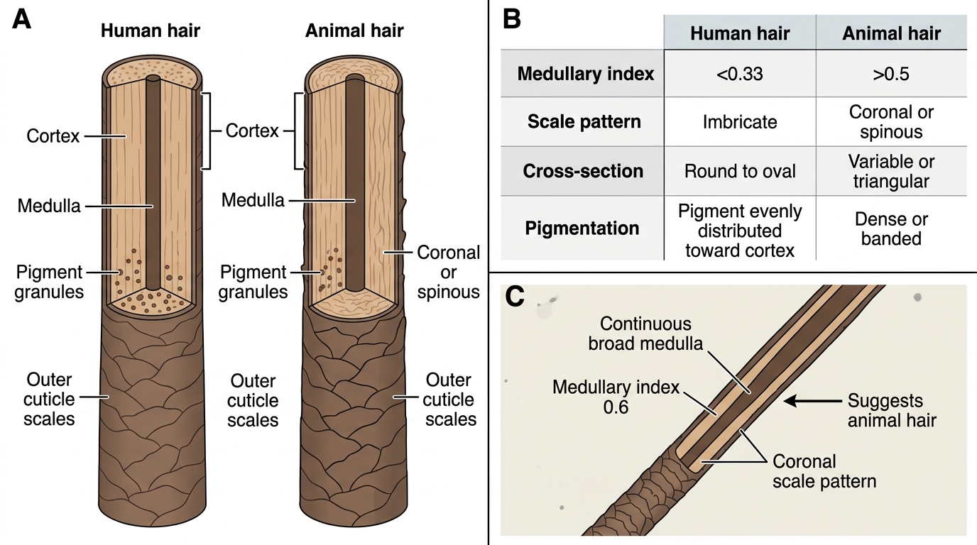
Microscopic Identification of Human vs Animal Hair
Fibre evidence is encountered in cases where contact between a suspect and victim can be evidenced by transfer of textile fibres. Natural fibres (cotton, wool, silk) and synthetic fibres (nylon, polyester, acrylic) each have characteristic microscopic cross-sections, birefringence patterns under polarised light, and chemical composition detectable by infrared spectroscopy. In the absence of a laboratory, the forensic doctor documents fibre colour, texture, and type and preserves them for forensic science laboratory analysis.
Anthropometry (Bertillon system): the historical precursor to fingerprinting, devised by Alphonse Bertillon in the 1880s, used 11 standardised body measurements to create a criminal identification card. It was abandoned after the 1901 Will West case at Leavenworth Federal Penitentiary, USA — where two men (Will West and William West) had nearly identical Bertillon measurements but completely different fingerprints. This case established the superiority of fingerprinting for individual identification. Anthropometry is now obsolete as an identification system but remains historically significant as the direct predecessor of modern dactylography.
SELF-CHECK
A hair is found at a crime scene. Under compound microscopy, it shows a continuous medulla with a medullary index of 0.6 and a coronal scale pattern. What is the MOST likely identification?
A. Human scalp hair
B. Human pubic hair
C. Animal hair
D. Synthetic fibre
Reveal Answer
Answer: C. Animal hair
A medullary index of 0.6 (>0.5) and coronal scale pattern are characteristic of animal hair. Human hair has a medullary index <0.33 and typically shows flattened/imbricate scale patterns. Pubic hair is still human and would show medullary index <0.33. Synthetic fibres are not biological and would not show a medulla or biological scale patterns.
Dactylography: Fingerprint Identification
Dactylography — the science of fingerprint identification — rests on two fundamental properties of friction ridge skin: permanence (ridge patterns form by the 4th month of fetal life and remain unchanged throughout a person's lifetime, barring deliberate destruction) and individuality (no two persons — including identical twins — have been shown to have identical fingerprint patterns across all ten digits). These properties make fingerprints the gold standard for criminal identification.
The three main fingerprint pattern types and their approximate prevalences in the general population (per Reddy's) are:
- Loops (~65%): the most common pattern; characterised by ridges that enter from one side, curve around, and exit from the same side. Further classified as ulnar loops (open toward the ulnar side of the hand — the side of the little finger) and radial loops (open toward the radial side — the thumb side). The loop pattern contains one triradius (delta).
- Whorls (~30%): ridges forming concentric circles or spiral patterns; contain two triradii. Four sub-types: plain whorl, central pocket loop whorl, double loop whorl, accidental whorl.
- Arches (~5%): the least common; ridges enter from one side and exit the other without curving back. Two sub-types: plain arch (smooth wave) and tented arch (a spike or tent-like ridge in the centre). Arches contain NO triradius.
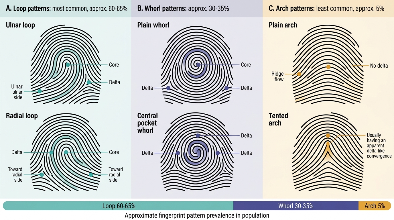
Major Fingerprint Pattern Types and Approximate Prevalence
The Henry Classification System (developed by Sir Edward Henry, introduced to India 1897 and to Scotland Yard 1901) provides the international standard for fingerprint filing and searching. It assigns each finger a numerical value based on whether it contains a whorl or not, generating a primary classification fraction that allows large fingerprint collections to be searched rapidly.
Ridge characteristics (minutiae) are the specific points where individual ridges bifurcate, end, or form islands — these are the comparison points used for individualisation. Common types include ridge ending, bifurcation, short ridge (enclosure), island (dot), and spur. Indian courts have accepted a minimum of 8–12 concordant minutiae with no unexplained discordances as the threshold for a positive fingerprint identification, though the standard varies by case and court; international standards range from 8 (Australia) to 16 (historically UK).
In post-mortem cases, fingerprints may be collected from decomposed or mummified bodies using specialised techniques: for macerated skin, the skin is removed from the finger, dried, and re-inked; for badly decomposed tissue, silicone casting or chemical enhancement methods are used. For latent fingerprints at crime scenes, development methods include powder dusting (aluminium powder for dark surfaces, carbon for light surfaces), ninhydrin (amino acid reaction, develops latent prints on paper), and cyanoacrylate fuming (on hard surfaces).