Page 9 of 15
FM4.4-5,FM14.8 | Identification from Skeletal & Other Remains — SDL Guide (Part 2)
Footprints, Poroscopy, and Other Surface Markers
Beyond fingerprints, several other surface features of the human body serve as individualising markers in forensic identification. Each has specific forensic applications, documentation methods, and levels of evidential strength.
Footprints are impressions left by the plantar surface of the foot. Like fingerprints, the friction ridges of the plantar surface are unique to the individual. Forensic footprint analysis considers: (1) the overall shape and dimensions of the foot impression (useful for general sizing — foot length correlates with stature); (2) the specific ridge detail of the toes and ball of the foot (as individualising as fingerprints, but less commonly recovered due to footwear); and (3) gait patterns (stride length, width, angle of step) in dynamic impressions in soft soil. Collection: photography with scale, casting in plaster or silicone for three-dimensional impressions.
Poroscopy is the study of the sweat pore pattern on friction ridge skin, developed by Edmond Locard in 1912. Each fingerprint ridge carries pores at regular intervals, but the position and shape of these pores on any given ridge are unique to the individual. Poroscopy provides a level of detail finer than ridge pattern comparison — it is used as confirmatory evidence when partial prints are available that show pores but insufficient ridge detail for standard minutiae comparison. The method requires high-magnification photography of lifted latent prints.
Scars are areas of fibrous tissue replacement following skin injury. Forensically, scars are permanent (unless surgically revised), individualising, and describable: shape, size, colour relative to surrounding skin, surface texture (raised/hypertrophic, depressed/atrophic, flat), and location. Scars may result from surgery, burns, accidents, or violence. Importantly, old scars may be the only surviving identifying feature when a body is partly decomposed. Documentation requires a standardised body diagram or photograph with scale measurement.
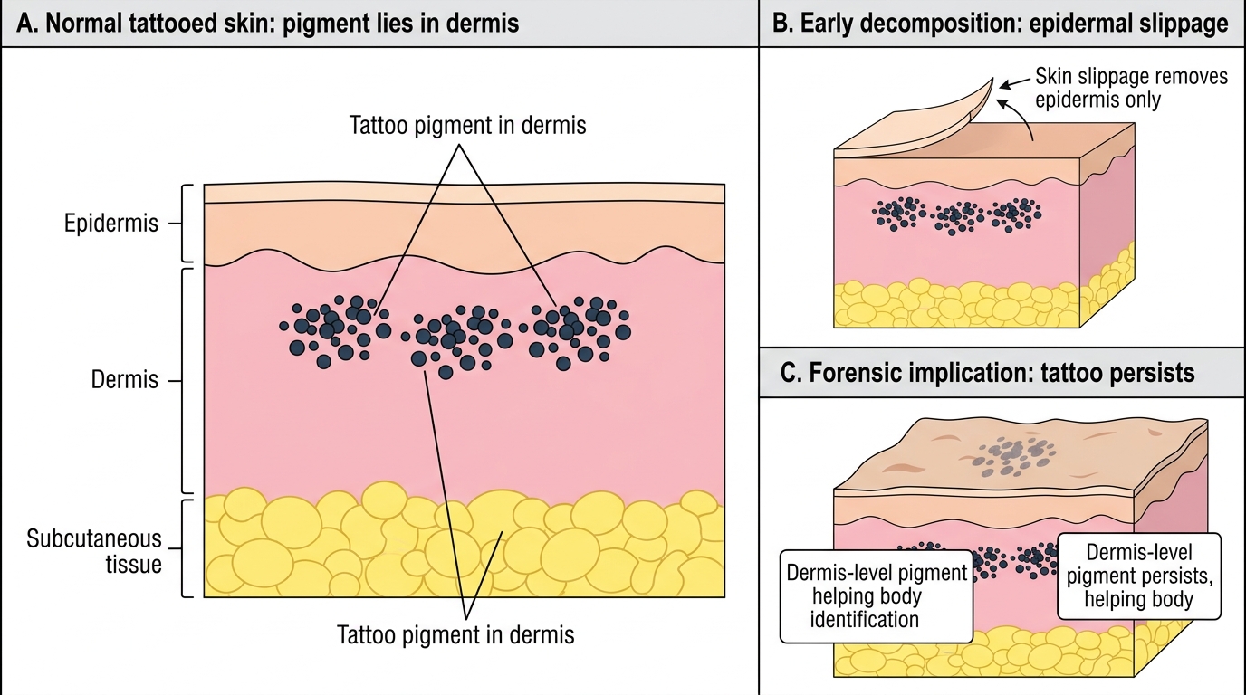
Tattoos are subdermal pigment deposits that are essentially permanent. Forensically, tattoos are documented by: design (symbolic, text, figurative, tribal), anatomical location, dimensions, colour, and professional versus amateur quality. Amateur tattoos use non-specialist pigments (carbon, plant-based inks) and show irregular edges and depth variation under dermoscopy; professional tattoos are more uniform. Tattoo databases are maintained by police departments in many countries and can identify bodies when other methods are unavailable. Crucially, tattoos survive mild decomposition because the pigment is in the dermis, not the epidermis — the epidermis may slough but the design often remains visible on the dermis.
Forensic Persistence of Tattoos During Early Decomposition
| Marker | Forensic Persistence | Individualisation Level | Primary Forensic Use |
|---|---|---|---|
| Fingerprint | Lifetime (until skin lost) | Very high (gold standard) | Criminal ID, victim ID |
| Footprint | Scene-dependent | High (if ridges visible) | Scene linkage, suspect exclusion |
| Scar | Lifetime (unless revised) | Moderate-high | Victim ID, suspect description matching |
| Tattoo | Lifetime (into decomposition) | Moderate-high | Victim ID, body identification |
| Poroscopy | As for fingerprint | Very high (supplementary) | Partial print confirmation |
SELF-CHECK
Which statement about dactylography and poroscopy is CORRECT?
A. Poroscopy compares the overall ridge pattern (loop, whorl, arch) between two prints
B. Dactylography refers to footprint analysis; poroscopy refers to fingerprint analysis
C. Poroscopy compares the pattern of sweat pores on friction ridge skin and provides finer individualisation detail than ridge pattern comparison
D. Arches are the most common fingerprint pattern, found in approximately 65% of the population
Reveal Answer
Answer: C. Poroscopy compares the pattern of sweat pores on friction ridge skin and provides finer individualisation detail than ridge pattern comparison
Poroscopy (Locard's method) compares the position and shape of sweat pores on friction ridge skin, providing a level of detail finer than minutiae comparison — it is used as confirmatory evidence for partial prints. Dactylography is fingerprint science (not footprint analysis). Loops are the most common pattern (~65%); arches are the least common (~5%). Overall ridge patterns (loop/whorl/arch) are the domain of the Henry classification, not poroscopy.
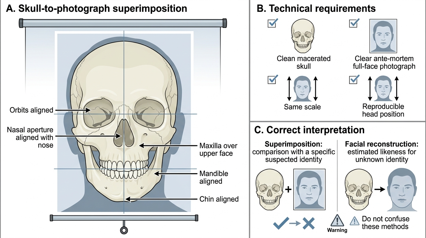
Superimposition: Skull and Photograph Comparison
Superimposition is a technique used to confirm or exclude the identity of an unknown skull by comparing it with an ante-mortem photograph of a known individual. It is a method of positive identification of a specific individual — and must be carefully distinguished from facial reconstruction (also called craniofacial reconstruction or facial approximation), which is a completely different technique.
The critical distinction that forensic doctors must maintain is: superimposition requires BOTH a known skull AND a known ante-mortem photograph of the same suspected person. The skull is positioned and scaled to match the angle, size, and orientation of the face in the photograph, and the two images are overlaid (traditionally using video cameras, now using digital image processing). A positive result requires that all skeletal landmarks (orbits, nasal bones, zygomatic arches, chin, ear canals) align precisely with the corresponding soft tissue features in the photograph. A single unexplained misalignment is grounds for exclusion. Superimposition can give a positive identification (the skull COULD be the person in the photograph) or an exclusion (the skull CANNOT be the person — a specific landmark does not match), but it cannot provide the same level of certainty as DNA or fingerprints.
Facial reconstruction, by contrast, works from a skull alone without any assumed identity — it builds up a probabilistic approximation of the living face using tissue depth data, muscle attachment points, and artistic technique. The goal is to generate a likeness that can be publicised to prompt recognition by the public, generating a lead for further investigation. It does NOT require a known photograph and does NOT produce a positive identification — it produces a lead.
Historically, superimposition was first used in the Ruxton case (1935, UK), where Buck Ruxton dismembered two bodies and the skulls of the victims were superimposed onto ante-mortem photographs, helping to identify the victims. In India, the technique has been applied in several high-profile murder cases.
Skull-to-Photograph Superimposition for Identification
Technical requirements for superimposition: (1) a clean, ideally macerated skull; (2) a clear ante-mortem photograph showing a full-face or near-full-face view; (3) the photograph and skull must be at the same scale; (4) the head position in the photograph must be reproducible on the skull. The technique is most useful when only one or two suspected identities exist — it is impractical as a screening tool across many candidates.
CLINICAL PEARL
The superimposition/facial reconstruction confusion is the single most common conceptual error in FM4.5 examinations. Students frequently write that superimposition 'reconstructs the face from the skull' — this is facial reconstruction, not superimposition. The forensic consequence is significant: superimposition gives a positive or negative comparison against a specific known person; facial reconstruction gives a probabilistic likeness for a complete unknown. In medicolegal reports and court testimony, confusing the two methods — especially claiming a positive identification by 'facial reconstruction' — would be professionally and legally untenable. Always state the method used and its specific requirements and limitations.
Medicolegal Inference: Report and Statutory Framework
The medicolegal reporting of skeletal remains and identification evidence must integrate scientific findings with statutory requirements and appropriate epistemic humility. The degree of certainty varies by method: DNA provides the highest certainty (random match probability stated as one in billions); fingerprints with 12+ concordant minutiae are accepted by Indian courts as positive identification; superimposition provides probable or excluded identification, not certainty; hair examination provides 'consistent with' or 'inconsistent with' language, not identification.
For the FM14.8 skill — examination and presentation of opinion on skeletal remains in a simulated or supervised environment — the standard examination sequence is:
- Document the scene of discovery (if attending) or note the source as reported
- Establish that remains are human (not animal) — compare skeletal anatomy with anatomical reference
- Establish number of individuals — count bones, look for duplicates (two right femora = minimum two individuals)
- Biological profile: sex (pelvis, skull), age (epiphyseal fusion, dental eruption or Gustafson, pubic symphysis), stature (Pearson's formula from long bones), race (skull morphology)
- Individualising features: any surviving attached soft tissue with scars, tattoos; ante-mortem injuries (old fractures with healed callus); post-mortem injuries or animal damage
- Dental charting for comparison with dental records
- Request forensic science laboratory examination of any associated trace evidence (hair, fibre)
- Estimate time since death from degree of decomposition, condition of bones, environment
The statutory framework for identity reporting:
- Indian Evidence Act Section 45: expert opinion on identification by the examining doctor is admissible as expert evidence
- Indian Evidence Act Section 73: fingerprints may be directed by court to be compared by an expert; court may itself compare prints
- Identification of Prisoners Act 1920: governs the collection of fingerprints (and photographs) from convicted and arrested persons; provides the legal basis for fingerprint databases
- CrPC Section 174: police authority to request PM examination of unidentified bodies
Report language for skeletal remains should explicitly state: findings from each parameter, method used, estimated biological profile with ranges, individualising features noted, what further laboratory tests are recommended, and what remains uncertain. A good medicolegal report on skeletal remains closes with: 'These findings are consistent with [profile]; final identification confirmation should be obtained by [dental record comparison / DNA profiling / fingerprint comparison] if ante-mortem records become available.'
Self-Assessment: Remains Identification
Test your understanding with the following questions before reviewing the answers.
Q1. A hair is submitted from a rape case. Microscopy shows: medullary index 0.28, flattened scale pattern, continuous cortex with fine distributed pigment. Is this human or animal hair? Explain your reasoning.
Q2. List the three main fingerprint patterns in order of prevalence (most to least common), with approximate percentages.
Q3. A detective asks you to 'reconstruct the face of this skull so we can identify the victim.' You instead propose superimposition. What is the key condition that must be satisfied before superimposition can be performed?
Q4. What does the Henry Classification System classify, and what is it used for?
Q5. A tattooed forearm is recovered from a river — the skin is partially macerated (skin slippage). Can the tattoo still be identified? Explain.
Answers:
1. Human hair — medullary index 0.28 (<0.33), flattened/imbricate scale pattern, and fine distributed cortical pigment are all characteristic of human hair. Animal hair would show medullary index ≥0.5 and coronal or spinous scale patterns.
2. (1) Loops — approximately 65%; (2) Whorls — approximately 30%; (3) Arches — approximately 5%.
3. Superimposition requires BOTH a known skull AND a known ante-mortem photograph of the suspected specific person. Without a suspected identity and ante-mortem photo, superimposition cannot be performed — what can be done instead is facial reconstruction.
4. The Henry Classification System classifies fingerprint patterns (primarily whether a digit contains a whorl or not) to generate a numerical fraction that enables large fingerprint collections to be searched and filed systematically. It underpins fingerprint bureau operations globally.
5. Yes — tattoo pigment is deposited in the dermis, not the epidermis. When the epidermis maceration (skin slippage) removes the outer layer, the tattoo design often remains visible on the dermis. The dermal design can be photographed and documented even after significant post-mortem change.