Page 3 of 18
MI4.1-4 | Diarrhoea & Dysentery — SDL Guide (Part 3)
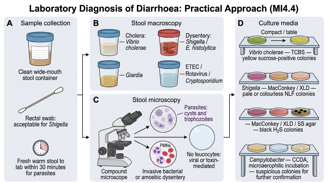
Laboratory Diagnosis of Diarrhoea — Practical Approach (MI4.4)
Laboratory Diagnosis of Diarrhoea: Practical Approach
Sample collection: Collect stool in a clean wide-mouth container. Rectal swabs acceptable for Shigella. For parasites: fresh warm stool within 30 minutes to the lab.
Macroscopy:
- Rice-water: cholera
- Bloody mucoid: dysentery (Shigella, E. histolytica)
- Fatty/greasy: Giardia
- Watery, non-bloody: ETEC, Rotavirus, Cryptosporidium
Microscopy: Wet mount (saline + iodine), concentration techniques (formol-ether)
- Cysts and trophozoites: parasites
- PMNs + RBCs: invasive bacterial/amoebic dysentery
- No leucocytes: viral or toxin-mediated
Culture media:
| Organism | Medium | Colony appearance |
|---|---|---|
| Vibrio cholerae | TCBS | Yellow (sucrose+) |
| Shigella | MacConkey, XLD | Pale/colourless (NLF) |
| Salmonella | MacConkey, XLD, SS agar | Black colonies (H₂S) on XLD/SS |
| Campylobacter | CCDA (microaerophilic) | Non-lactose fermenter, comma rods |
Rapid tests: Cholera dipstick (Crystal VC), Rotavirus antigen ELISA, C. difficile toxin EIA

Stool Appearance to Microscopy Algorithm for Diarrhoea
CLINICAL PEARL
Leucocyte count in stool predicts invasion: In bacillary dysentery (Shigella), the wet mount shows sheets of PMNs with RBCs. In amoebic dysentery, there are fewer PMNs (trophozoites secrete leucolytic factors) but erythrophagocytosis is seen. In cholera or viral diarrhoea, leucocytes are absent. This single observation guides empirical treatment before culture results return.
SELF-CHECK
In the stool microscopy of a patient with dysentery, you identify motile trophozoites with ingested red blood cells. Which of the following additional features would confirm Entamoeba histolytica rather than non-pathogenic species?
A. Presence of chromatoid bodies in cysts
B. Erythrophagocytosis by the trophozoite
C. Quadrinucleate cyst on iodine mount
D. Absence of PMNs in the stool
Reveal Answer
Answer: B. Erythrophagocytosis by the trophozoite
Erythrophagocytosis — the ingestion of red blood cells by trophozoites — is the pathognomonic finding distinguishing Entamoeba histolytica from E. dispar and other Entamoeba species. Chromatoid bodies and quadrinucleate cysts are seen in both E. histolytica and E. dispar (morphologically identical). Absence of PMNs is a relative finding in amoebiasis but is not confirmatory.