Page 1 of 12
MI1.1-2 | Introduction to Microbiology & Bacterial Morphology — SDL Guide
Learning Objectives
- Describe the historical milestones and key scientists who shaped medical microbiology
- Explain the role of microbes in maintaining health and causing disease
- Classify bacteria, viruses, fungi, and parasites using basic taxonomic criteria
- Describe the morphology, physiology, and characteristic features of major microbial groups
- Enumerate common infections caused by representative organisms in each microbial category
INSTRUCTIONS
Medical microbiology underpins your understanding of infectious disease — the leading cause of mortality in India. This module grounds you in the discipline's historical foundations and gives you the classificatory framework that every subsequent microbiology topic builds upon. Engage with the micro-quizzes to check recall as you read.
References
- Ananthanarayan & Paniker's Textbook of Microbiology, 11th ed., Ch 1-2, Ch 4 (textbook)
- Jawetz, Melnick & Adelberg's Medical Microbiology, 28th ed., Ch 1 (textbook)
Version 2.0 | NMC CBUC 2024
CLINICAL SCENARIO
In 1854, London's Broad Street pump handle was removed — and a cholera epidemic stopped almost overnight. John Snow, a physician without a microscope, used geographic mapping alone to implicate contaminated water. Twenty years later, Robert Koch would prove what Snow suspected: invisible living agents cause specific diseases. Today you carry that same investigative inheritance every time you order a culture or interpret a Gram stain.
WHY THIS MATTERS
Understanding microbial classification and morphology is not academic taxonomy — it directly drives clinical decision-making. A Gram-positive coccus in clusters points toward Staphylococcus and dictates empiric anti-staphylococcal cover; a Gram-negative rod from a urinary specimen narrows the differential to Enterobacteriaceae. Without this foundational map, antibiotic choices become guesswork.
RECALL
From Year 1 Biochemistry, you know that cell membranes are phospholipid bilayers and that proteins are polymers of amino acids. From Physiology, you are familiar with the concept of homeostasis and host defence. Recall that DNA encodes proteins and that replication errors drive variation — these same principles apply to microbial genetics. Keep these in mind as you learn how bacteria differ from human cells, and why that difference enables selective antimicrobial toxicity.
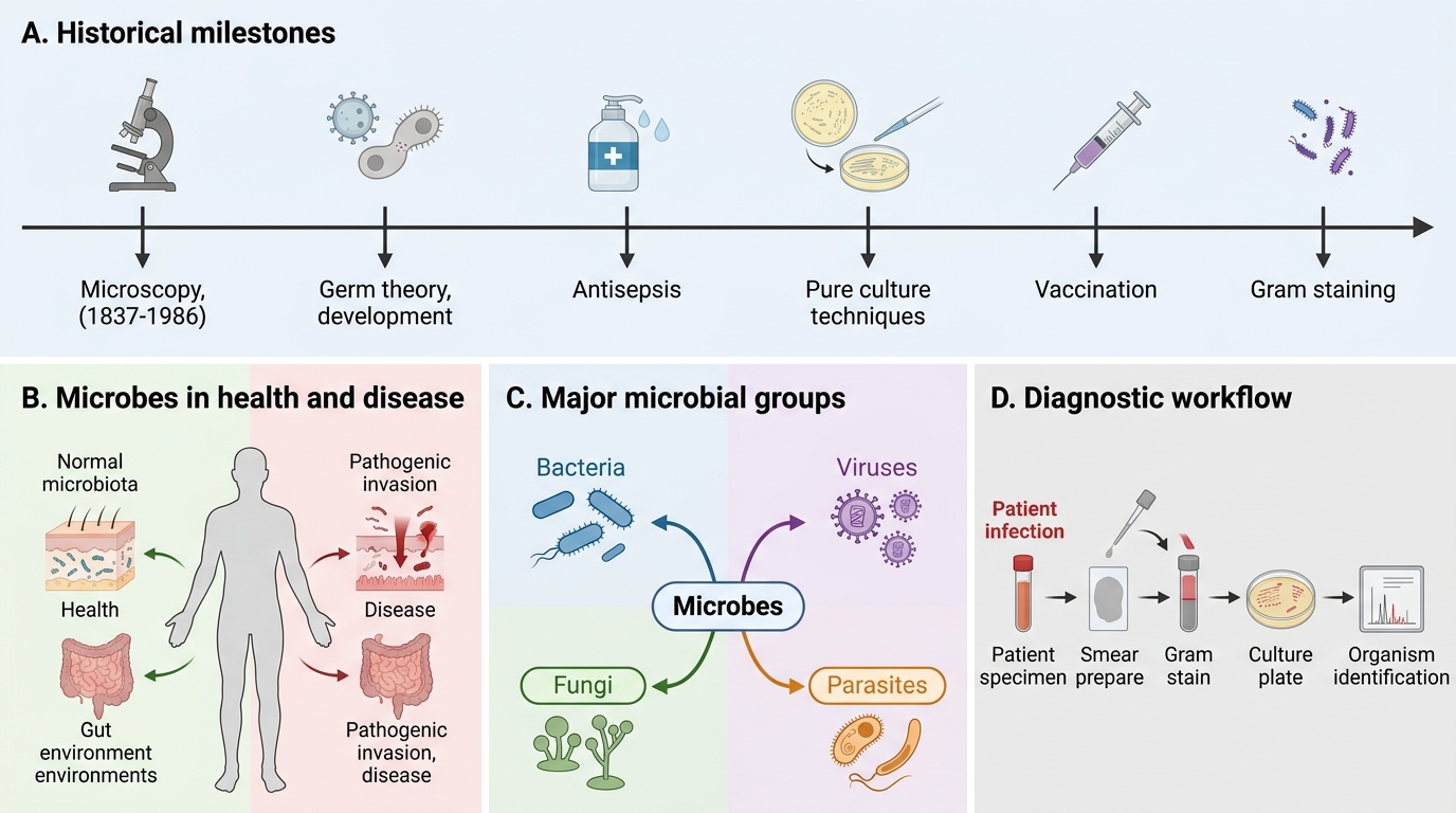
Historical Milestones in Medical Microbiology
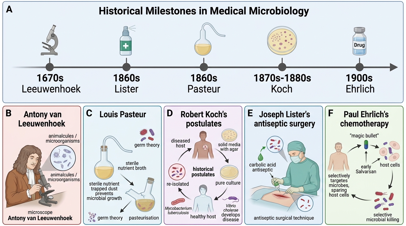
Historical Milestones in Medical Microbiology
The modern discipline rests on a series of conceptual revolutions:
Antony van Leeuwenhoek (1670s) first visualised animalcules (now called microorganisms) using his simple microscope — establishing that an invisible living world exists.
Louis Pasteur (1860s) disproved spontaneous generation with his swan-neck flask experiment and developed germ theory: specific microbes cause specific diseases. He also pioneered vaccines (cholera, anthrax, rabies) and pasteurisation.
Robert Koch (1870s-80s) formalised the causal relationship using Koch's postulates:
1. The organism must be found in all cases of the disease.
2. It must be isolated in pure culture from the host.
3. The pure culture must reproduce the disease in a healthy host.
4. The organism must be re-isolated from the experimentally infected host.
Koch also developed solid media (agar, thanks to Angelina Fanny Hesse's suggestion), identified Mycobacterium tuberculosis (1882) and Vibrio cholerae (1883), and received the Nobel Prize in 1905.
Joseph Lister (1860s) introduced antiseptic surgical technique using carbolic acid, dramatically reducing post-operative mortality — a direct clinical application of germ theory.
Paul Ehrlich (1900s) developed the concept of chemotherapy: searching for a 'magic bullet' that kills microbes without harming the host. His compound Salvarsan (arsphenamine) became the first specific antimicrobial agent (against syphilis, 1909).
Alexander Fleming (1928) observed that Penicillium mould lysed Staphylococcus colonies — leading ultimately to penicillin and the antibiotic era.
Indian contributions: Sir Ronald Ross (working in India, 1897) demonstrated that malaria is transmitted by the Anopheles mosquito — earning the Nobel Prize in 1902. Waldemar Haffkine pioneered cholera and plague vaccines in India in the 1890s.

Milestones in General Microbiology
Koch's postulates have been modified for obligate intracellular parasites (e.g., viruses) and for organisms identified by molecular methods alone (molecular Koch's postulates, Falkow 1988).
Role of Microbes in Health and Disease
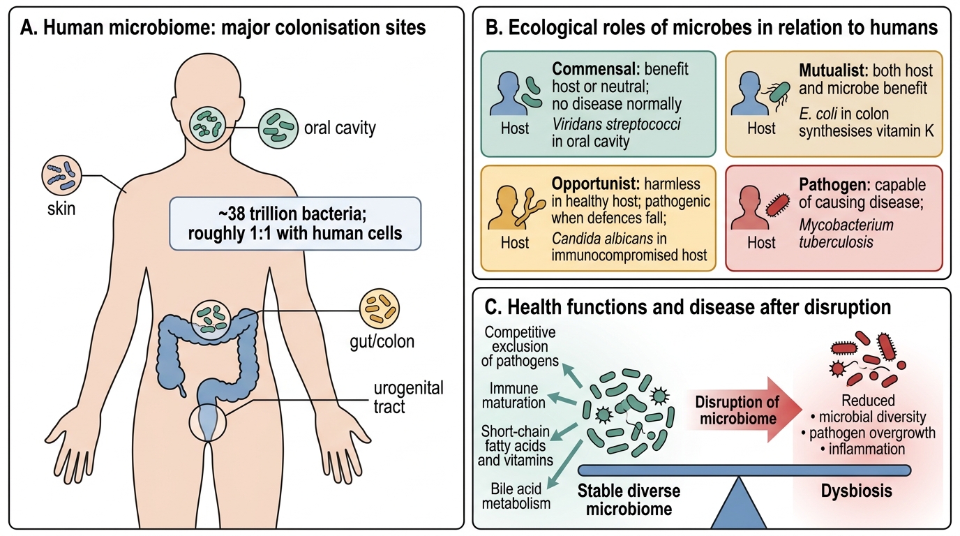
Role of Microbes in Health and Disease
Microorganisms occupy four ecological roles in relation to humans:
| Role | Definition | Example |
|---|---|---|
| Commensal | Benefit host or are neutral; cause no disease under normal conditions | Viridans streptococci in oral cavity |
| Mutualist | Both host and microbe benefit | E. coli in colon synthesises Vitamin K |
| Opportunist | Harmless in healthy hosts; pathogenic when host defences fall | Candida albicans in immunocompromised |
| Pathogen | Always capable of causing disease | Mycobacterium tuberculosis |
The human microbiome comprises ~38 trillion bacteria (roughly 1:1 with human cells) colonising skin, gut, oral cavity, and urogenital tract. Functions include:
• Competitive exclusion of pathogens
• Maturation of innate and adaptive immunity
• Synthesis of short-chain fatty acids and vitamins
• Metabolism of bile acids
Disruption of the microbiome (dysbiosis) — by antibiotics, illness, or diet — is linked to Clostridioides difficile colitis, inflammatory bowel disease, and metabolic syndrome.
Pathogenicity is the capacity to cause disease; virulence quantifies the degree. Key virulence determinants include:
• Adhesins — surface molecules that facilitate attachment to host cells
• Invasins — enable penetration of epithelial barriers
• Toxins — exotoxins (secreted proteins) and endotoxins (LPS of Gram-negative bacteria)
• Capsules — resist phagocytosis (Streptococcus pneumoniae, Klebsiella)
• Biofilm formation — protects from antibiotics and immune clearance
SELF-CHECK
Koch isolated Mycobacterium tuberculosis in 1882. Which of his postulates requires that the isolated organism must reproduce the disease when inoculated into a healthy, susceptible host?
A. Postulate 1 — organism found in all cases
B. Postulate 2 — organism isolated in pure culture
C. Postulate 3 — pure culture reproduces the disease
D. Postulate 4 — organism re-isolated from experimental host
Reveal Answer
Answer: C. Postulate 3 — pure culture reproduces the disease
Postulate 3 establishes causality by demonstrating that the purified organism alone is sufficient to reproduce the disease in a healthy host. This distinguishes a true pathogen from a mere bystander. Postulate 4 closes the loop by re-isolating the same organism.
Classification of Microorganisms
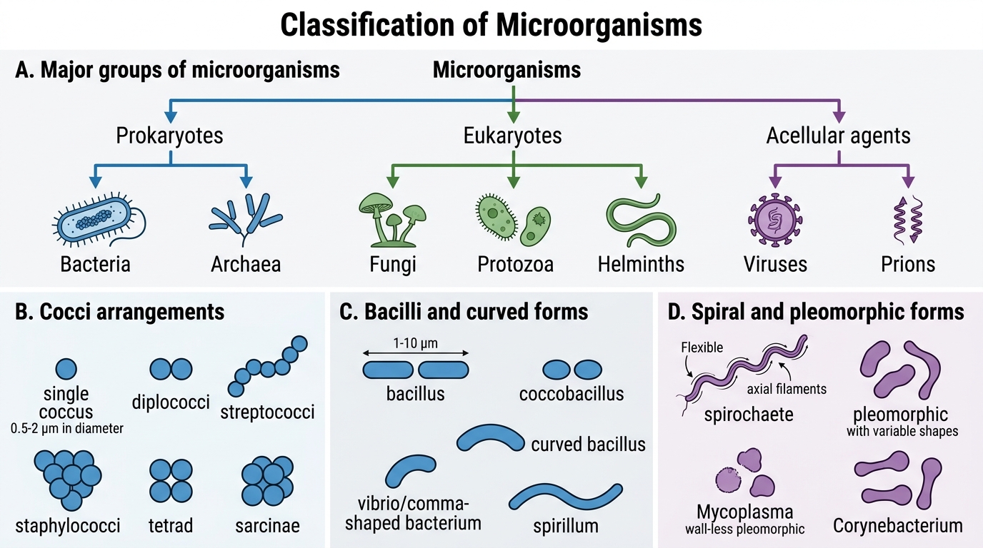
Classification and Morphology of Microorganisms
The microbial world is divided into two fundamental cell plans:
Prokaryotes (no membrane-bound nucleus): Bacteria and Archaea
Eukaryotes (membrane-bound nucleus): Fungi, Protozoa, Helminths
Acellular agents (not classified as living by strict criteria): Viruses, Prions
### Bacteria
Classification by shape:
• Cocci — spherical (diameter 0.5-2 µm): arranged as singles, pairs (diplococci), chains (streptococci), clusters (staphylococci), tetrads, packets of eight (sarcinae)
• Bacilli — rods (1-10 µm length): straight (E. coli), curved (vibrios), comma-shaped
• Spirilla — rigid spirals (Campylobacter)
• Spirochaetes — flexible spirals with axial filaments (Treponema, Leptospira)
• Pleomorphic — variable shape (Mycoplasma, Corynebacterium)

Bacterial Morphologies and Cocci Arrangements
Classification by Gram stain:
• Gram-positive — thick peptidoglycan (20-80 nm), retain crystal violet → appear purple
• Gram-negative — thin peptidoglycan (2-7 nm) + outer membrane, decolourised → appear red/pink with safranin counterstain
• Gram-variable / Gram-indeterminate — Mycobacteria (acid-fast), Mycoplasma (no cell wall)
Bacterial structure:
• Cell wall — provides shape; composed of peptidoglycan (murein) — the target of β-lactam antibiotics
• Cytoplasmic membrane — semi-permeable bilayer; site of oxidative phosphorylation (no mitochondria)
• Ribosomes — 70S (30S + 50S subunits) — distinct from human 80S ribosomes; targeted by aminoglycosides, tetracyclines, macrolides, chloramphenicol
• Nucleoid — single circular dsDNA chromosome, without histone proteins
• Plasmids — extrachromosomal circular DNA; carry antibiotic resistance and virulence genes
• Capsule (some) — anti-phagocytic polysaccharide layer
• Flagella (some) — locomotive organelle; composed of flagellin protein
• Pili/Fimbriae — attachment (fimbriae) and conjugation (sex pili)
• Spores (Clostridium, Bacillus) — dormant forms resistant to heat, desiccation, chemicals

Bacterial Cell Structure
Viruses, Fungi, and Parasites — Overview
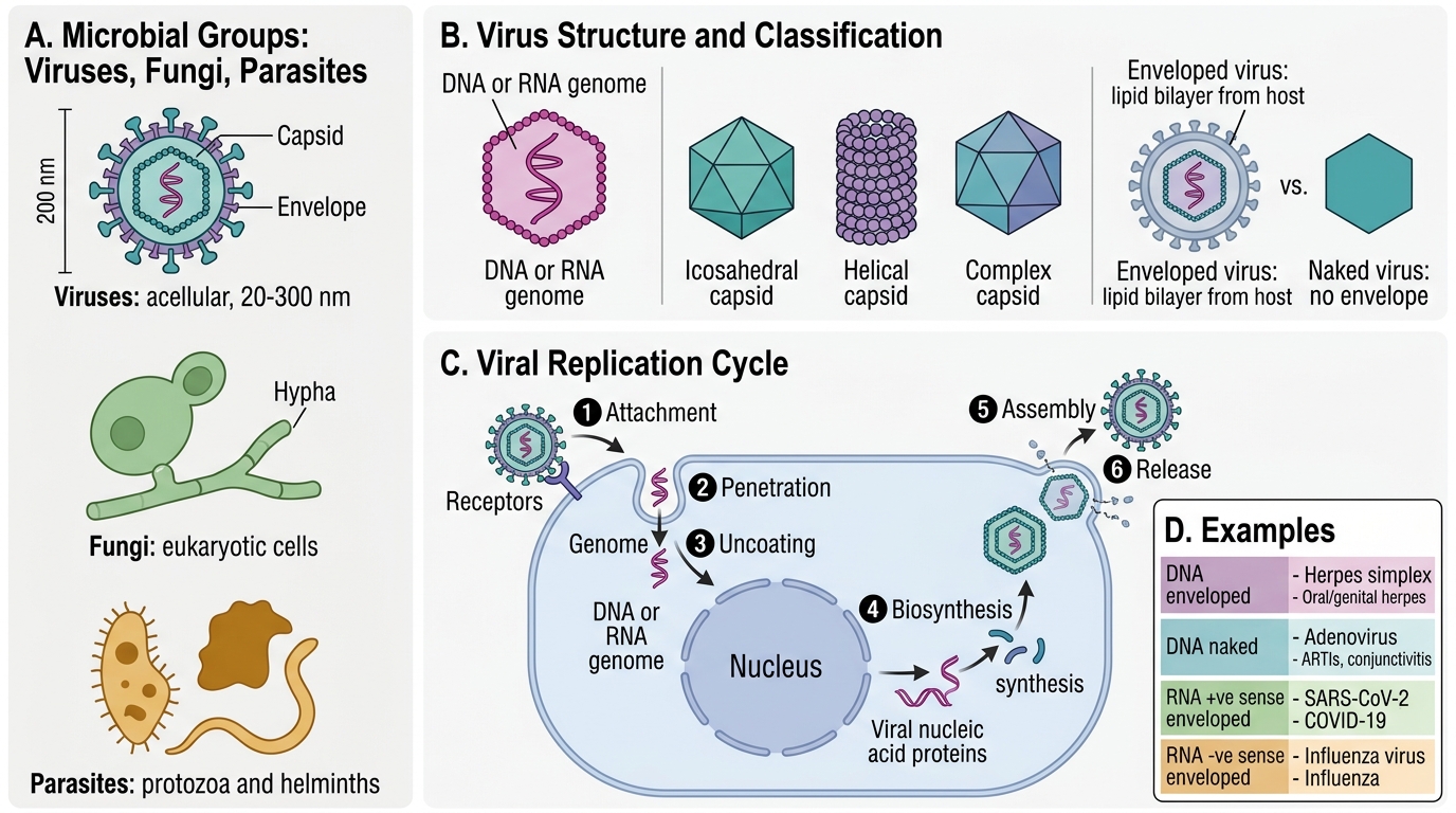
Overview of Viruses, Fungi, and Parasites
### Viruses
Viruses are acellular obligate intracellular parasites, 20-300 nm in size. They lack ribosomes and metabolic machinery; they hijack host cell machinery for replication.
Classification:
• By nucleic acid: DNA (single- or double-stranded) vs RNA (single- or double-stranded, +ve or −ve sense)
• By capsid symmetry: icosahedral, helical, complex
• By envelope: enveloped (lipid bilayer derived from host, e.g., HIV, Influenza) vs naked/non-enveloped (e.g., Poliovirus, Hepatitis A)
Viral replication cycle: Attachment → Penetration → Uncoating → Biosynthesis → Assembly → Release
| Virus type | Example | Disease |
|---|---|---|
| DNA, enveloped | Herpes simplex | Oral/genital herpes |
| DNA, naked | Adenovirus | ARTIs, conjunctivitis |
| RNA, +ve sense, enveloped | SARS-CoV-2 | COVID-19 |
| RNA, −ve sense, enveloped | Influenza A | Seasonal flu |
| Retrovirus | HIV | AIDS |
### Fungi
Fungi are eukaryotes with ergosterol-containing cell membranes (target of antifungals) and chitin-containing cell walls (absent in bacteria and human cells).
Forms:
• Yeasts — unicellular, reproduce by budding (Candida, Cryptococcus)
• Moulds — multicellular filamentous, form hyphae and mycelium (Aspergillus, Rhizopus)
• Dimorphic fungi — yeast in tissue (37°C), mould in environment (25°C): Histoplasma, Blastomyces, Sporothrix
Common infections: candidiasis, dermatophytosis (ringworm), cryptococcal meningitis, aspergillosis (especially in immunocompromised patients).
### Parasites
Parasites include protozoa (unicellular eukaryotes) and helminths (multicellular worms):
Protozoa — Plasmodium (malaria), Entamoeba histolytica (amoebic dysentery), Giardia lamblia (giardiasis), Leishmania (kala-azar)
Helminths — nematodes (roundworms: Ascaris, hookworm), cestodes (tapeworms: Taenia), trematodes (flukes: Schistosoma)
India bears a disproportionate global burden of malaria, kala-azar (visceral leishmaniasis), lymphatic filariasis, and soil-transmitted helminthiases.
CLINICAL PEARL
When you see 'sepsis with Gram-positive cocci in clusters' in a blood culture report, think Staphylococcus aureus first — particularly in IV drug users, post-surgical patients, or those with indwelling lines. However, in neonates, Gram-positive cocci in chains suggest Streptococcus agalactiae (Group B Strep) rather than staph. The arrangement pattern is a rapid triage tool while sensitivities are pending.
SELF-CHECK
A 28-year-old woman with HIV (CD4 count 45 cells/µL) presents with fever and headache. India ink preparation of CSF shows encapsulated yeasts. The causative organism belongs to which microbial category?
A. Bacterium (Gram-positive)
B. Virus (DNA, enveloped)
C. Fungus (yeast form)
D. Protozoan (intracellular)
Reveal Answer
Answer: C. Fungus (yeast form)
Cryptococcus neoformans is a yeast-form fungus with a polysaccharide capsule that appears as a clear halo around the yeast cells on India ink staining. It is a classic opportunistic pathogen in advanced HIV disease (CD4 <100). India ink is not used for bacteria or viruses; it is specific for encapsulated fungi in CSF analysis.
REFLECT
Think about the last news report you heard about an infectious disease outbreak — COVID-19, mpox, drug-resistant TB, or dengue. Using the framework in this module, can you classify the causative agent and explain why it is pathogenic? Consider: what virulence factors does it possess? Is it an obligate intracellular pathogen, or can it replicate extracellularly? What host defence mechanisms does it evade? This habit of mapping clinical observations onto microbial biology is the core skill of the clinical microbiologist.
KEY TAKEAWAYS
Medical microbiology evolved from van Leeuwenhoek's microscope through Pasteur's germ theory to Koch's postulates, establishing that specific microbes cause specific diseases. Microorganisms occupy commensal, mutualist, opportunistic, or pathogenic roles; virulence depends on adhesins, invasins, toxins, and immune evasion.
Microbial classification distinguishes:
• Bacteria (prokaryotes) — classified by shape, Gram reaction, and structural features; cell wall peptidoglycan and 70S ribosomes are antibiotic targets
• Viruses (acellular) — obligate intracellular; classified by nucleic acid type, capsid symmetry, and envelope
• Fungi (eukaryotes) — yeasts, moulds, dimorphic; ergosterol and chitin distinguish them from host cells
• Parasites — protozoa and helminths; major burden in India
Morphological identification on Gram stain remains a cornerstone of early clinical decision-making.