Page 4 of 12
MI1.10 | Staining Techniques — Gram & ZN (Practical) — SDL Guide (Part 2)
Ziehl-Neelsen (ZN) Acid-Fast Staining — Principle and Procedure
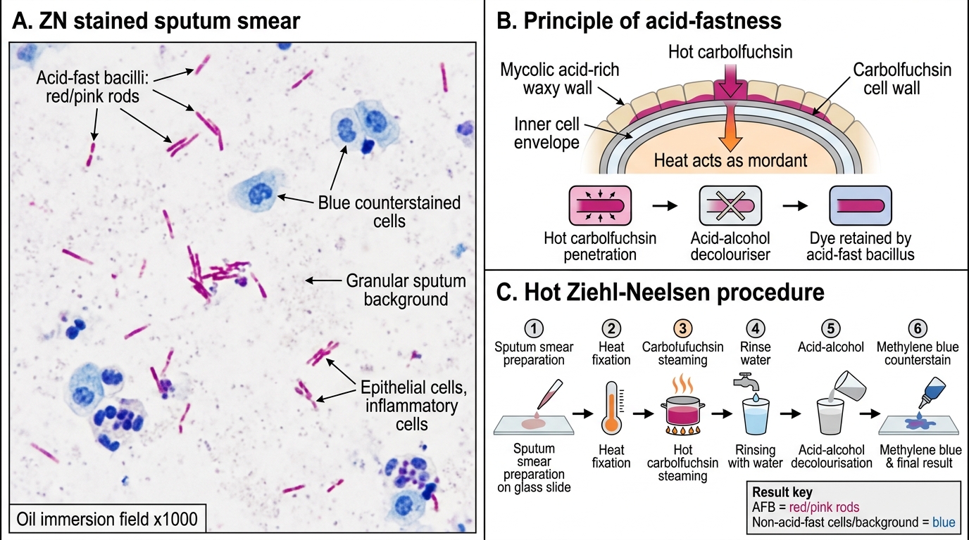
Ziehl-Neelsen Acid-Fast Staining: Principle and Procedure
Mycobacteria possess a waxy, mycolic acid-rich cell wall that resists penetration by aqueous stains. The ZN stain overcomes this resistance using heat and concentrated carbolfuchsin; once stained, mycobacteria resist decolourisation even with acid-alcohol (hence 'acid-fast').
Principle:
1. Carbolfuchsin (hot) — heat drives the phenol-fuchsin dye through the mycolic acid layer into the cell. Heat = the mordant.
2. Acid-alcohol (3% HCl in 70% alcohol — decolouriser) — removes carbolfuchsin from non-acid-fast organisms and background; mycobacteria retain the red dye due to their waxy wall.
3. Methylene blue (counterstain) — stains non-acid-fast organisms and background cells blue.
Result:
• Acid-fast bacilli (AFB) → red/pink (magenta) rods
• Non-acid-fast organisms, cells → blue

Ziehl-Neelsen Stained Sputum Smear: AFB Identification
Procedure (hot ZN/Ziehl-Neelsen method):
1. Prepare a smear from the most purulent/caseous part of sputum; air-dry, heat-fix.
2. Cover with carbolfuchsin; heat gently from below (steam, not boil) for 5 minutes — replenish dye if it evaporates; do not allow to dry.
3. Cool, then rinse with water.
4. Decolourise with acid-alcohol (3% HCl in 70% alcohol) until no more red colour washes off (~1-2 min).
5. Rinse with water.
6. Apply methylene blue counterstain for 30-60 seconds; rinse and blot dry.
7. Examine under oil immersion (×1000).
Cold ZN method (Kinyoun's): uses higher concentration of carbolfuchsin with a wetting agent; no heating required. Used for Cryptosporidium and Cyclospora oocysts (modified ZN).
Reporting under NTEP (National Tuberculosis Elimination Programme):
| Grade | AFB count | Report |
|---|---|---|
| Negative | 0 AFB in 100 fields | No AFB seen |
| Scanty (1+) | 1-9 AFB per 100 fields | Specify exact count |
| 1+ | 10-99 AFB per 100 fields | Positive |
| 2+ | 1-10 AFB per field (in 50 fields) | Positive |
| 3+ | >10 AFB per field (in 20 fields) | Positive |
At least 100 oil immersion fields must be examined before reporting as negative.
CLINICAL PEARL
India follows the NTEP sputum grading scale for ZN smears — know these categories. A 'scanty' result (1-9 AFB in 100 fields) requires exact enumeration and clinical correlation; it does not automatically mean TB (could be a NTM or environmental contamination). In a patient on anti-TB therapy, a still-positive ZN smear at 2 months is a key decision point for treatment modification. The ZN smear also identifies Cryptosporidium parvum oocysts (modified cold ZN) — especially important in HIV patients with chronic diarrhoea.
Routine Stool Examination — Principle and Procedure
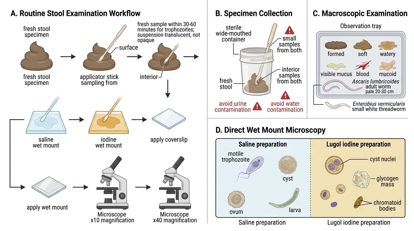
Routine Stool Examination: Principle and Procedure
Routine stool examination (RSE) identifies intestinal parasites — protozoa, cysts, ova, larvae, and adult worms — and is complemented by direct microscopy.
Specimen collection:
• Fresh stool sample, not more than 30-60 minutes old for trophozoite detection.
• Collect from different parts of the specimen (surface, interior) using an applicator stick.
• Volume: ~2-5 g (pea-sized portion).
• Avoid contamination with urine or water (destroys trophozoites).
Macroscopic examination:
Note: consistency (formed, soft, watery, mucoid), colour, blood or mucus, adult worms visible to naked eye (Ascaris — pale, ~20-30 cm; Enterobius — small white threadworms at perineum).
Microscopic examination — direct wet mount:
1. Place a drop of normal saline (0.9% NaCl) on one half of a clean slide.
2. Place a drop of Lugol's iodine on the other half.
3. Using an applicator stick, emulsify a small amount of stool in each drop — the suspension should be translucent (not opaque).
4. Cover with a coverslip; examine under ×10, then ×40 (high dry) objective.
Saline preparation: visualises trophozoites (motile), cysts, ova, larvae.
Iodine preparation: stains glycogen and nuclei — improves visualisation of cyst nuclei and chromatoid bodies.
| Parasite | Key microscopic feature |
|---|---|
| Entamoeba histolytica (trophozoite) | Directional pseudopodal motility, ingested RBCs in cytoplasm |
| Entamoeba histolytica (cyst) | 1-4 nuclei, chromatoid bars, glycogen mass |
| Giardia lamblia (trophozoite) | Pear-shaped, 2 nuclei, falling-leaf motility |
| Giardia lamblia (cyst) | Oval, 4 nuclei |
| Ascaris lumbricoides (ovum) | Oval, thick shell, mammillated outer layer |
| Hookworm (ovum) | Oval, thin shell, 4-8 cell stage |
| Trichuris trichiura (ovum) | Barrel/football shaped, bipolar plugs |

Diagnostic Stool Microscopy: Protozoa and Helminth Ova
Concentration techniques (increase diagnostic yield):
• Formol-ether (Ridley) concentration: centrifugation sediment examined — used when direct smear is negative
• Flotation (ZnSO₄, saturated NaCl): cysts and ova float; used for nematode ova
SELF-CHECK
On routine stool examination of a 5-year-old with diarrhoea, you see oval cysts with 4 nuclei in the iodine preparation. The child's mother says she noticed the child passes pale bulky stools. What is the most likely organism?
A. Entamoeba histolytica cyst
B. Giardia lamblia cyst
C. Cryptosporidium oocyst
D. Balantidium coli cyst
Reveal Answer
Answer: B. Giardia lamblia cyst
Giardia lamblia cysts are oval with 4 nuclei visible on iodine staining — a classic examination finding. Giardiasis causes malabsorption syndrome with pale, bulky, fatty (steatorrhoeic) stools due to villous atrophy and disruption of fat absorption in the small intestine. E. histolytica cysts have 1-4 nuclei but typically cause dysentery (bloody stools). Cryptosporidium oocysts are much smaller (4-6 µm) and require modified ZN staining.