Page 8 of 33
OP4.3 | Infective Keratitis: Differential Diagnosis and Management — SDL Guide (Part 2)
HSV Dendritic Keratitis: Diagnosis and Treatment
Herpes simplex virus (HSV-1, rarely HSV-2) causes the most common viral keratitis in clinical practice. Following primary oral infection in childhood, HSV becomes latent in the trigeminal ganglion and can reactivate to affect the cornea — often triggered by fever, UV exposure, stress, or immunosuppression. Recurrent HSV keratitis is a leading cause of corneal scarring and keratoplasty in developed countries.
The classic presentation is the dendritic ulcer: a branching, tree-like (dendritic = tree-like) epithelial lesion with characteristic terminal bulbs at the tips of each branch. These bulbs distinguish a true HSV dendritic ulcer from a healing epithelial defect (which has tapered ends, no bulbs) and from other viral keratitides. The ulcer stains brilliantly with both fluorescein (which pools in the epithelial defect) and rose bengal (which stains devitalised and virally infected epithelial cells surrounding the lesion — including the perimeter of the dendrite where the virus is actively replicating). This dual staining pattern is a clinical key: most other corneal ulcers stain predominantly with fluorescein; rose bengal positivity at the ulcer margins is a marker of viable virally infected cells.
As the disease progresses, the dendritic pattern may coalesce into a larger, irregular geographic (amoeboid) ulcer — a serious complication. The epithelium is lost over a large area. Geographic ulcers develop when topical steroids are applied without antiviral cover, when the diagnosis is missed, or when the patient is immunocompromised. This is the critical treatment trap for HSV keratitis: topical corticosteroids in active epithelial HSV keratitis cause the dendritic to expand into a geographic ulcer and massively worsen the prognosis. Students must memorise this.
Deep herpetic disease: in stromal (interstitial) HSV keratitis (covered partially in SDL 2), the epithelium may be intact but there is stromal oedema and disciform opacity. In this form, controlled use of steroids WITH antiviral cover is appropriate and anti-inflammatory treatment may be needed. The distinction between epithelial keratitis (antivirals alone; steroids contraindicated) and stromal keratitis (antivirals + carefully titrated steroids) is critical.
Treatment of epithelial HSV dendritic keratitis:
- Topical aciclovir 3% ointment five times daily for 14 days — this is the standard treatment
- Topical ganciclovir 0.15% gel (5 times daily) is an equally effective alternative with better penetration
- Oral aciclovir 400 mg five times daily (or valaciclovir 500 mg twice daily) is used for severe disease, immunocompromised patients, or recurrent disease (long-term prophylaxis)
- Mechanical débridement (gentle wiping of the infected epithelium with a cotton bud) speeds healing by removing virally laden cells — can be combined with antiviral drops
- Cycloplegics for associated uveitis
- NEVER use topical steroids without antiviral cover in active epithelial disease
Sensory reduction: HSV keratitis causes progressive reduction of corneal sensation with each recurrence (due to axonal damage of the long ciliary nerves by the virus). Testing corneal sensation with a cotton wisp is therefore diagnostically useful — markedly reduced sensation in a patient with recurrent eye disease strongly suggests HSV keratitis or neurotrophic keratitis.
Acanthamoeba Keratitis: Contact Lens Risk and Management
Acanthamoeba keratitis is caused by a free-living amoeba found in soil, freshwater, tap water, and swimming pools. It is almost exclusively a disease of contact-lens wearers, particularly those who rinse lenses with tap water, use homemade saline, swim or shower while wearing contact lenses, or sleep in lenses. Acanthamoeba exists in two life-cycle forms: the actively feeding trophozoite and the environmentally resistant double-walled cyst. When introduced onto a contact-lens-traumatised corneal surface, the trophozoite invades the epithelium and stroma, and — crucially — demonstrates a unique tropism for corneal nerve fibres. This neurotropism is the mechanistic basis for the hallmark clinical feature of Acanthamoeba keratitis: pain that is dramatically out of proportion to the visible inflammatory signs. Whereas bacterial keratitis produces severe pain that matches the degree of stromal destruction, in Acanthamoeba the pain is neuritic — originating from amoebic invasion of stromal nerve sheaths — and can be excruciating even when the slit-lamp findings appear relatively mild. Understanding this mechanism allows the clinician to hold the diagnosis in mind even before the classic slit-lamp signs appear, which is essential because early diagnosis dramatically improves outcome. The organisms colonise the contact lens storage case and are introduced onto the corneal surface where minor epithelial trauma from the lens allows invasion — making lens hygiene the central preventable risk factor.
The clinical hallmarks of Acanthamoeba keratitis make it one of the most distinctive — and most commonly misdiagnosed — corneal infections:
- Severe pain out of proportion to the degree of clinical signs — the disproportionate intensity of pain is the single most important diagnostic clue. The pain is neuritic in quality because Acanthamoeba has a predilection for invading corneal nerves.
- Radial keratoneuritis — pathognomonic, if present: radial infiltrates following the course of corneal stromal nerves, visible on slit-lamp as white lines radiating from the centre. This represents amoebic infiltration along the neural sheaths.
- Ring infiltrate — in established disease, a characteristic ring-shaped stromal infiltrate develops around the central cornea (the immune reaction surrounding the central focus of infection). The ring may be complete or partial.
- Minimal initial surface epithelial defect — early in the disease the epithelium may be relatively intact (or show only punctate keratitis), which can mislead the clinician into thinking the condition is mild.
Diagnosis: corneal scraping with Giemsa or calcofluor white staining to identify Acanthamoeba cysts (double-walled, polygonal). Culture on non-nutrient agar layered with E. coli as the food source. Confocal microscopy can demonstrate cysts in vivo.
Treatment: Acanthamoeba keratitis is notoriously difficult to treat and requires prolonged therapy — often 3–12 months:
- Polyhexamethylene biguanide (PHMB) 0.02% eyedrops hourly + Chlorhexidine 0.02% eyedrops hourly (biguanide combination) — these are cysticidal
- Propamidine isethionate 0.1% (Brolene) + neomycin as adjuncts in some protocols
- Topical steroids are generally avoided in early disease but may be used later under specialist supervision for severe stromal reaction
- Pain management is an important adjunct given the severity of neuritic pain
- Prognosis is poor if diagnosis is delayed — corneal perforation can result in advanced disease requiring keratoplasty
SELF-CHECK
A 26-year-old contact-lens wearer presents with excruciating right eye pain, described as burning and neuritic, for 2 weeks. Slit-lamp shows radial white infiltrates along the course of corneal stromal nerves. She reports rinsing her contact lenses with tap water. Which organism is most likely, and which culture medium is specific?
A. Pseudomonas aeruginosa; blood agar
B. Fusarium species; Sabouraud's dextrose agar
C. Acanthamoeba species; non-nutrient agar with E. coli overlay
D. Herpes simplex virus; no culture medium needed (clinical diagnosis)
Reveal Answer
Answer: C. Acanthamoeba species; non-nutrient agar with E. coli overlay
Radial keratoneuritis (radial infiltrates following nerve tracks) combined with contact-lens use, tap-water exposure, and severe neuritic pain are pathognomonic of Acanthamoeba keratitis. Acanthamoeba is cultured on non-nutrient agar with E. coli overlay — the E. coli serves as the food source for the amoeba. Blood agar is for bacteria; Sabouraud's for fungi; HSV is diagnosed clinically (dendritic morphology with rose bengal/fluorescein staining).
Differential Diagnosis Framework and Investigation
The bedside differential diagnosis of corneal ulcer is one of the most important clinical reasoning exercises in ophthalmology. It requires integrating the history (onset speed, trauma type, contact lens history, systemic illness) with the slit-lamp appearance (infiltrate character, margins, satellite lesions, staining pattern) to arrive at the most likely organism category BEFORE culture results are available — because treatment must often begin empirically.
The diagnostic framework is best structured around the key distinguishing features of each type:
Bacterial keratitis: rapid onset (24–48 hours), contact lens history (Pseudomonas) or trauma/surface disease; dense creamy-white infiltrate with ill-defined margins and surrounding stromal oedema; moderate to large hypopyon; mucopurulent discharge. The infiltrate grows rapidly. Gram stain from scraping is typically positive within hours.
Fungal keratitis: subacute onset (days to 1–2 weeks); vegetative trauma history (agricultural, rural); dry grey-white infiltrate with feathery/hyphate margins; satellite lesions; endothelial plaque; thicker viscous hypopyon. KOH mount reveals hyphae (septate for Aspergillus/Fusarium; non-septate for Mucor — very rare corneal pathogen).
HSV dendritic keratitis: branching dendritic ulcer with terminal bulbs; stains with both fluorescein AND rose bengal; history of recurrence or prior labial herpes; reduced corneal sensation; NO hypopyon (unless there is secondary bacterial superinfection). Progression to geographic ulcer if treated with steroids alone.
Acanthamoeba keratitis: contact-lens wearer + tap water exposure; severe neuritic pain out of proportion to signs; radial keratoneuritis; ring infiltrate. Giemsa stain or calcofluor white of scraping → double-walled cysts.
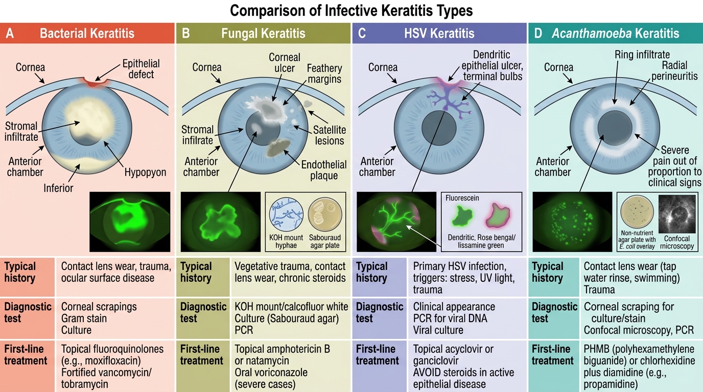
Comparison of Infective Keratitis Types
| Type | History clue | Slit-lamp appearance | Staining | Culture medium | First-line treatment |
|---|---|---|---|---|---|
| Bacterial | Contact lens; rapid | Dense creamy infiltrate; hypopyon | Fluorescein (epithelial defect) | Blood agar, chocolate agar | Fluoroquinolone or fortified antibiotics |
| Fungal | Vegetative trauma; rural | Feathery margins; satellite lesions; endothelial plaque | Fluorescein (defect); KOH mount (+) | Sabouraud's agar | Natamycin 5% |
| HSV | Recurrence; lip herpes; prior episodes | Dendritic with terminal bulbs | Fluorescein AND rose bengal | No culture (clinical); PCR | Topical aciclovir 3% or ganciclovir 0.15% |
| Acanthamoeba | Contact lens + tap water | Ring infiltrate; radial keratoneuritis | Giemsa/calcofluor white (cysts) | Non-nutrient agar + E. coli | PHMB + chlorhexidine |
Empirical treatment decision: When culture results are pending, the history and slit-lamp pattern guide empirical therapy. In rural India with vegetative trauma → start natamycin empirically (covers fungal) while awaiting KOH mount. In urban contact-lens wearer with rapid onset → fluoroquinolone empirically (covers bacterial) while awaiting Gram stain. If ANY dendrite-like pattern is seen → add topical aciclovir. Never withhold treatment for 24+ hours while awaiting culture in a rapidly progressing ulcer.
CLINICAL PEARL
The single most dangerous clinical error in corneal ulcer management is giving a bacterial antibiotic to a patient with HSV dendritic keratitis, while also adding a steroid 'to reduce inflammation.' The steroid without antiviral cover causes the dendritic ulcer to expand into a massive geographic ulcer. Before prescribing a steroid for any red eye with corneal involvement, ask: have I excluded HSV epithelial keratitis? If you can see a branching epithelial lesion with terminal bulbs, the answer is antivirals first, no steroids until the epithelial disease is controlled. A second equally dangerous error: treating a fungal ulcer with antibiotics and corticosteroids (the typical empirical cocktail prescribed when the vegetative trauma history is missed), which accelerates fungal invasion.